Bioimaging research
Bioscience Technology Facility
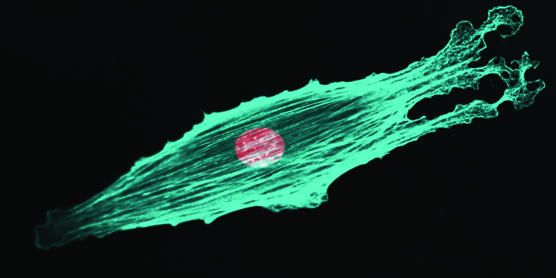
The Bioscience Technology Facility is pioneering new optical imaging modalities for its users and pushing the frontiers of optical superresolution microscopy, including collaborations with Phase Focus and Carl Zeiss. The facility constantly strives to improve its capabilities and explore new methods in support of the world-leading research at York.
Read more about the new Bioscience Technology Facility.

Peter O'Toole
Research Title: Director of the Bioscience Technology Facility and Head of Imaging and Cytometry in the Department of Biology.
Peter has a particular interest in combining different “omics” modalities to better understand cellular phenotypes.
The York Neuroimaging Centre (YNiC)
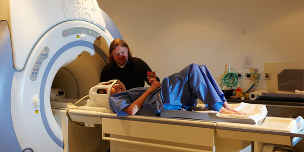
Understanding the workings of the human brain has fascinated researchers for centuries. Magnetic resonance techniques such as magnetic resonance imaging (MRI) and Magnetoencephalography (MEG) affords new and novel insights into the function of the brain in its entirety.
The York Neuroimaging Centre boasts two of the most powerful brain scanners in the UK that allow detailed study of a wide range of brain functions and conditions. Read more about the York Neuroimaging Centre.
Tony Morland
Research Title: Professor and co-Director of York Neuroimaging Centre.
Tony’s interest is in understanding how the brain deals with deficits in visual information that arise from disease or damage to the visual system.
Alex Wade
Research Title: Professor and co-Director of York Neuroimaging Centre.
Alex’s interest is in understanding neurological disease and the perception of colour.
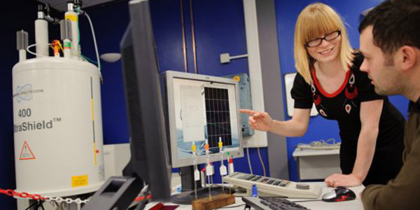
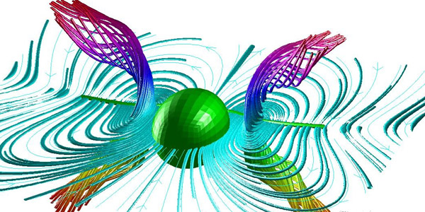
Centre for Hyperpolarisation in Magnetic Resonance (ChyM)
The Centre for Hyperpolarisation in Magnetic Resonance (ChyM) aims to explore the fundamental processes that underpin hyperpolarisation techniques. In particular, CHyM tackles fundamental sensitivity issues associated with nuclear magnetic resonance (NMR) spectroscopy and magnetic resonance imaging (MRI).
Read more about the Centre for Hyperpolarisation in Magnetic Resonance.

Simon Duckett
Research Title: Professor and Director of the Centre for Hyperpolarisation in Magnetic Resonance.
Professor Duckett's interest is in NMR-related techniques, such as parahydrogen induced polarisation methods to study chemical reactions or hyperpolarisation methods to sensitise biological substrates.
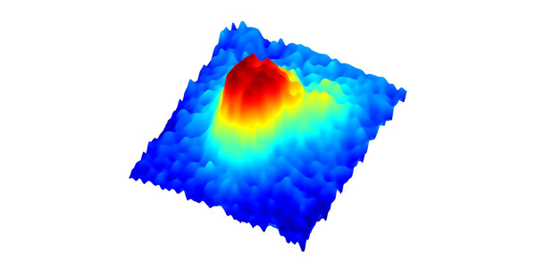
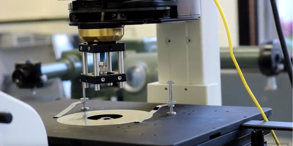
Resonant Hyperspectral Imaging
Being able to image individual proteins without the need for fluorescent labels is one of the holy grails in biomedical imaging. Researchers at York have developed a novel hyperspectral imaging method that exploits nanophotonic resonances to make the tiniest particles visible. They have recently used this technique to answer fundamental questions in cell secretion and cell signalling.
Find out more about Resonant Hyperspectral Imaging.
Thomas F Krauss
Research Title: Professor of Photonics.
Professor Krauss is a world-leading expert in photonic nanostructures, with a key interest in developing novel nanophotonic concepts for addressing biological questions.
Single-molecule cellular biophysics
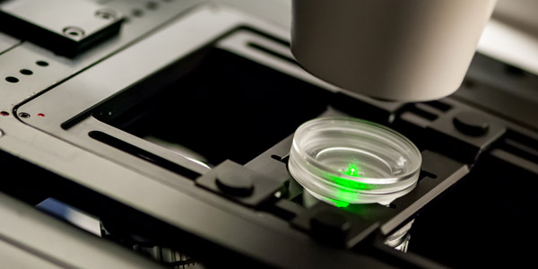
Single-molecule cellular biophysics specialises in single molecule fluorescence imaging investigations on living cells to explore and understand the foundations of life at the molecular level. The group develops bionanotechnology tools for application in biomedicine, synthetic biology and bioengineering.
Read more about the single-molecule cellular biophysics research.

Mark Leake
Research Title: Professor Anniversary Chair of Biological Physics.
Professor Leake has a special interest in developing and applying novel forms of optical microscopy to investigate complex biological processes at the level of single molecules.
Holographic microscopy
Optical imaging is typically confined to a two-dimensional image plane. In order to understand the motion of cells in their native three-dimensional environment, we need to develop new imaging methods. Borrowing concepts from holographic imaging, holographic microscopy aims to capture the movements of cells in 3D space and in real time in order to better understand their behaviour.
Find out more about Holographic microscopy research.
Laurence Wilson
Research Title: Lecturer.
Dr Wilson has developed a novel holographic imaging method that allows the three-dimensional study of microorganisms such as bacteria and their movement in real time.
Mathematical Biology and Chemistry
Mathematical Biology and Chemistry develops cutting-edge mathematical approaches for studying challenging problems, especially in image analysis. The group studies biological fluid dynamics, such as swimming sperm and bacteria, mathematical ecology; such as fisheries and stochastic dynamics, mathematical physiology relevant to wound healing and systems biology, and a number of related topics.
Read more about our Mathematical Biology research.
Julie Wilson
Research Title: Reader with a joint appointment between the Departments of Mathematics and Chemistry.
Dr Wilson has a key interest in the application of mathematical and statistical methods to biological and chemical problems, such as novel methods for image analysis.