Our impact
Explore case studies from researchers working with communities around the world towards positive change.
Posted on 29 January 2018
Our biologists are at the forefront of new developments in scientific imaging
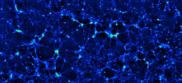
Neuronal cells seen under a microscope using ptychographic imaging
The potential applications for this technology are immense...”
Biologists at York are at the centre of a major advance in microscope technology which opens up important new insights into cell structure and behaviour.
Researchers worked with Sheffield-based scientific imaging specialists Phasefocus on the development of a microscope which greatly improves the clarity of cell images.
The breakthrough relies on an imaging method known as ptychography. A major benefit of the technology is that it allows scientists to study cells without the need to stain the samples with markers, such as fluorescent labels or dyes to make them easier to view.
Markers and dyes affect the structure and function of living cells and can disrupt processes such as cell movement and division.
“Looking through the new microscope is like watching cells on High Definition TV in terms of the clarity, contrast and precision of the images,” said Dr Peter O’Toole, Director of the Bioscience Technology Facility and Head of the University’s Imaging and Cytometry Laboratory.
“We can see subtle changes in cellular features, shape and movement. These new insights allow us to watch and understand the living behaviour of cells.”
Label-free ptychography also provides researchers with important quantitative information such as cell size, mass and volume.
The first commercial version of the new Livecyte microscope was launched in Spring 2017. At least six systems have already been sold, including one to our Department of Biology to complement the beta-test version being used by researchers across the department.
Dr O’Toole, who has been leading the application work at York, said it was an important breakthrough in scientific imaging technology with many potential research applications including cancer research, pharmaceutical development and stem cell research.
“The potential applications for this technology are immense – most areas of biology that use primary cells from living tissue are potential benefactors.”
The project has resulted in five research papers involving biologists at York who have highlighted unique applications for the new microscope.
The microscope was developed by Phasefocus assisted by an Innovate UK and EPSRC-funded Knowledge Transfer Partnership (KTP) grant – a government scheme which allows businesses to grow by linking them to a university and a graduate to work on a specific project.
In this case, the KTP funded the recruitment of biologist Dr Rakesh Suman who worked on the development of applications for the new technology and validation of a prototype microscope in York’s Biology laboratories.
Dr O’Toole said: “This project is a perfect example of how academic institutions can work with industry to develop commercial applications for our world class research and laboratory facilities.
“As a University, we are a perfect test bed for this type of technology. We have the laboratory infrastructure and importantly, we also have access to specialists such as cancer research biologists, neurobiologists, immunologists, stem cell specialists. Not only can these researchers test out the technology – but they can also use it to help them answer important research questions.”
He added: “This project is about really exciting science, but it’s also about working with industry to develop commercial applications for our expertise. It’s a mutually beneficial arrangement.”
Dr Suman continues to work on the project at the Imaging and Cytometry labs at York, but he is now employed full time by Phasefocus. He has been joined by Dr Richard Kasprowicz, a former PhD student at York who is also now employed by the company.
Dr Martin Humphry, Chief Executive Officer of Phasefocus said: “This arrangement means we have scientists embedded in a working department with access to researchers with real biological questions to be answered. The feedback resulting from our partnership has helped shape the development of our Livecyte instrument and identify issues that really matter to active researchers.”
Researchers at York, led by Dr O’Toole are now exploring new applications for the microscope technology. Some of the University’s key biology research teams are already working with the new product.
Dr O’Toole explained: “Our partnership with Phasefocus, has proved that the technology works, now we want to use label-free ptychography to help us explore some of our most pressing research challenges.”
The text of this article is licensed under a Creative Commons Licence. You're free to republish it, as long as you link back to this page and credit us.

Dr Peter O’Toole
Research interests in fluorescence imaging and flow cytometry
Label-free imaging to study phenotypic behavioural traits of cells in complex co-cultures was published in Nature Scientific Reports
Ptychography – a label free, high-contrast imaging technique for live cells using quantitative phase information was published in Nature Scientific Reports
Characterising live cell behaviour: Traditional label-free and quantitative phase imaging approaches was published in The International Journal of Biochemistry & Cell Biology
Find out more about the work of Phasefocus
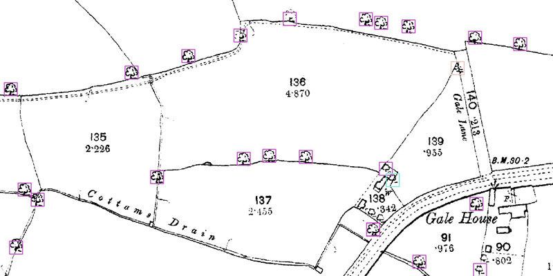
A research project needed to spot trees on historic ordnance survey maps, so colleagues in computer science found a solution.
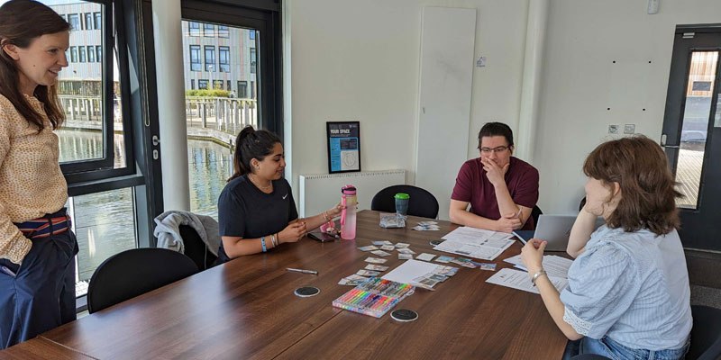
We’re using gaming technology to ensure prospective teachers are fully prepared for their careers.
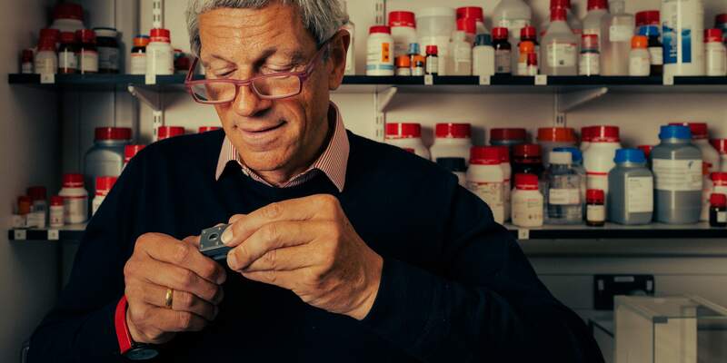
A low cost, high-accuracy device, could play a large part in the NHS's 'virtual wards'.
Our monthly research newsletter features a curated mix of news, events, and recent discoveries delivered straight to your inbox.