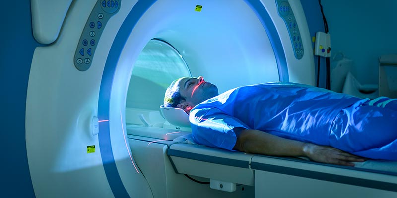
Functional magnetic resonance imaging (fMRI)
Our projects

Combining virtual reality with fMRI and multivariate pattern analyses to reveal the neural representations that support spatial navigation.

Combining experimental psychology, computational modelling, and brain imaging to reveal how forgetting differs across brain regions.
Examining how the brain sees 3D motion by comparing signals from the left and right eyes.

Exploring how monolinguals and bilinguals produce the words that they want to say.

Examining how different types of distraction are ignored and how this relates to working memory capacity.

Using computational modelling of light penetration to explore its effects on the brain.
Measuring the anatomical and physiological changes in the eyes and brain associated with increased peripheral visual sensitivity in deaf adults.

Using fMRI to explore developmental dyscalculia and dyslexia.

Exploring whether reading comprehension and production of sentences recruit similar brain mechanisms and connectivity patterns.

Using fMRI to measure patterns of neural response to objects and faces in the visual brain.