
New PET scan technique will be able to improve cancer treatment
Posted on Tuesday 15 September 2026
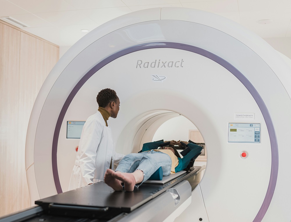
PET scans are a standard technological tool for finding and observing diseased human tissue. It relies on the analysis of high energy (gamma) photons emitted by a tracer that is injected into a patient's body, but it is both time-consuming and prone to errors due to motion blur.
Researchers from institutions in Tokyo and York are collaborating to refine this technology using laboratory detector arrays.
3D model
During PET scans, patients are injected with a tiny amount of, for example, radioactively labelled sugar. This is because rapidly dividing cancer cells consume massive amounts of sugar to fuel their growth, making the sugar accumulate around the tumors and acting as a tracer to locate the cancerous cells.
A computer software is then able to create a 3D model of the tracer’s locations by analysing the trajectories of millions of photons that the tracer emits in pairs when it decays. As this software measuring two photons needs time and millions of inputs to create the 3D image, the current PET scan technology is sensitive to motion blur, often arising when the patient breathes etc.
Three photons
While two photons is the standard outcome, occasionally three photons are emitted and by calculating their positions and trajectories, the researchers from Tokyo and York were able to successfully image and spatially resolve the relative contribution of this three-photon process, for the first time.
Whether two or three photons are created is highly dependent on its immediate chemical environment, particularly on the concentration of local oxygen. By also measuring the ratio of three-photon to two-photon, this new model and technique will allow doctors and scientists to create a detailed oxygen map of the scanned tissue.
Tumors that are low in oxygen are notoriously resistant to standard radiation therapy. With this new technique, doctors could not only locate a tumor but also determine whether it is oxygen-deficient. This extra context allows medical teams to choose better alternative treatments from the start and monitor how well a tumor responds over time.
Implications
With these models it will be possible to more easily pinpoint the affected tissue in real time, making it possible to continuously study both how the diseased tissue behaves as well as the effectiveness of any treatment. The new three-photon model will be able to pave the way for more precise, personalised patient care and has major implications for cancer treatment.
The research was carried out by scientists from the University of York, University of Tokyo and Institute Of Science Tokyo, Positron Emission Tomography.
The full paper was published in Communications Physics: Advancing PET through direct imaging of three-photon decay using pure positron emitters | Communications Physics