
Tying the knot: scientists reveal hidden mechanism controlling viral replication
Posted on Thursday 3 September 2026
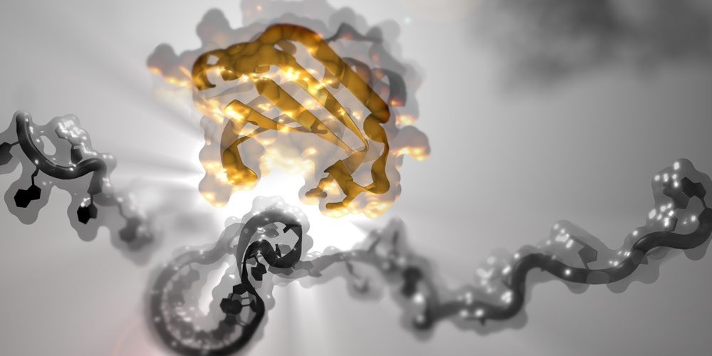
Researchers in the York Structural Biology Laboratory and the Department of Biology have led an international research collaboration in virology. Together with colleagues from the Physics of Life group, University of Sheffield, University of Cambridge and EMBL Hamburg, they have discovered how sections of a virus genome change shape to control whether viral proteins are made or not.
The findings, which are published in Molecular Cell coincide with the 50th anniversary of the launch of YSBL in 1976.
Dr Chris Hill explains that a large number of ribonucleic acid (RNA) viruses, including SARS-Cov-2 and HIV-1, use a mechanism known as ‘frameshifting’ to make their proteins, and if that mechanism can be disrupted, it could be possible to stop the virus from replicating.
He said: “Basically, the ribosome; the protein-making machine, ‘slips’ at a particular place while moving along the viruses' genetic code, usually when it encounters a ‘knot’ in the RNA. This isn't a random error, it's a carefully orchestrated event - if we stop it happening the viruses are severely compromised.
“Crucially, we know this ‘slipping’ rarely happens during protein synthesis in uninfected cells, so if we could understand it at a molecular level we might be able to find ways of stopping it from happening, which could lead to new antiviral drugs.”
Dr Hill says that while this ‘slipping’ mechanism is well-known to science, his team have managed to capture images which give an entirely new understanding of these processes.
“In this paper, we show that one specific group of viruses, cardioviruses, actually use a viral protein to force the RNA into a knot only when the protein is present. We've managed to take photos using X-ray crystallography and Small-Angle X-ray Scattering (SAXS) of exactly how this protein causes the knot to be tied.
“We combined this with sophisticated fluorescence microscopy, one molecule at a time, to observe the RNA as it flips from ‘unknotted’ to ‘knotted’. This is a completely new mechanism in gene expression.”
Dr Hill says it’s possible that many other proteins found in the cell might interact with these ‘knots’ and his team is now planning more research to find out.
“It’s a really exciting discovery. It adds to a growing body of evidence that RNA isn't just an information-storage molecule, but that its precise 3D shape - knotted versus unknotted - is vitally important at controlling how genes are turned on and off - regulation that is fundamental to life.”