Our equipment and services
Flow Cytometer
Rapid single cell analysis and sorting. For both routine testing and further detailed analysis of both simple and complex, difficult to handle sample types. Expertise ranges from rare cell analysis and sorting, simultaneous cell cycle and receptor analysis, FRET, receptor quantification, small particle (e.g. EV’s and bacteria) analysis, through to large plant protoplasts!
Our labs consist of multiple flow cytometers to enable most types of flow cytometry. They are all housed in temperature controlled Cat II rooms and can pipeline to any of the ‘omics core labs.
Cytometers include:
- Beckman Coulter MoFlo Astrios with 4 laser lines (405, 488, 561 and 633 nm). This system is capable of 6-way (separating and collecting 6 different populations simultaneously), sterile sorting in excess of 80,000 cells per second and sorting into up to 1535 well plates.
- Beckman Coulter CytoFLEX SRT with 4 lasers, 15 colour detectors and 384 well plate sorting. This is a an easy, walk up and use sorter for most routine sample types.
- 2 x Beckman Coulter CytoFLEX LX with 6 lasers ranging from 355 nm to 808 nm and 21 colour detectors. All CytoFLEX systems can utilise single tube or multiwell plate loading.
- Becton Dickinson Fortessa X-20 with 4 lasers and 16 colour detectors.
- Beckman Coulter CytoFLEX S with 4 lasers and 15 colour detectors.
Contact us to find out more
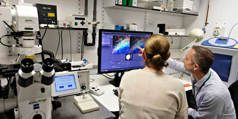
Advanced light microscopy
We have expertise and cover a wide array of most light microscopy techniques from standard confocal to super resolution techniques with expertise in many biophysical applications, such as FCS, FCCS, FRAP, FRET, PALM, STORM, SIM and much more. We also have great expertise in many sample types, ranging from the classical cultured cells to human tissues, bacteria, yeast, algae, plant sample types through to many material samples.
Our advanced light microscopy area consists of different confocal and multiphoton microscopes, super resolution microscopes through to label-free technologies with the aim to cover as many techniques as possible. All microscopes are in temperature controlled Cat II labs with technical help always nearby. The labs are well equipped and available to both internal and external users.
Microscopes (with incubation) include:
- Carl Zeiss ELYRA 7 (for PALM/STORM/SIM2 live cell imaging). This is a 4 laser two camera based system.
- Carl Zeiss LSM980 MP AiryScan 2. This system includes multiphoton for in vivo and deeper imaging. This can be used for simple through to complex confocal and multiphoton applications and also has LSM Plus to enable resolutions down to close to 100 nm. This also has the spectral head as standard, as well as the non-descanned detectors for the multiphoton.
- Carl Zeiss LSM880 AiryScan and LSM780 confocal microscopes come on an invert and upright microscope respectively. These cover lasers lines from 405 though to 633 nm and both have full spectral detector capabilities.
Electron microscopy
For sub-micron and even sub-nanometre resolution of various sample types, the transmission electron microscope (TEM) is extensively used for both biological and chemical studies of cell sections and novel compounds.
The scanning electron microscope (SEM) is used for morphological studies of even more varied sample types. These vary from single cells to light bulb elements.
There is a range of peripheral equipment.
Our TEM is an FEI Tecnai 12 G2 fitted with a CCD camera for ease and speed of use. The resolution is around 0.4nm and capable of discriminating small variation in size of nm particles. The TEM is also used to study protein localisation and co-localisation with gold particles. Our technician has all the necessary experience to prepare samples of varying kinds (plant and animal sections, immuno-labelling of cryosections, nanoparticles samples).
The SEM is ideal for detailed morphological studies which are beyond the resolution of light microscopes. Samples vary from the monocultured cell to plant leaves, light bulb filaments through to metal chips.
Equipment
- TEM - FEI Tecnai 12 G2
- 20kV-120kV high voltage range BioTWIN configuration for high contrast imaging
- Computer-controlled high stability Compustage
- Single and multigrid specimen holders
- Fully embedded digital imaging system with SIS Megaview III camera
- Transmission electron microscope
- SEM - JEOL JSM-6490LV
- High and low vacuum capability
- Secondary and backscattered electron imaging
- Quorum Technologies PP2000 cryosystem and low vacuum capabilities
- Scanning electron microscope
Contact us to find out more
Analysis software packages
We have a range of off-line analysis packages for image analysis and flow data analysis:
- Software
- Image Analysis
- Volocity (server access required - please ask a member of I&C to register you)
Ptychography
Label-free Imaging
We are innovators and early adopters of quantitative phase imaging techniques, these include, Ptychography for prolonged time-lapse and screening in multi well plates through to digital holography for rapid 3D super resolution, label-free imaging.
Ptychography is a novel means of label-free, high-contrast live cell imaging for easy cell tracking and growth analysis, even through plastic cell culture dishes. The high-contrast artefact free images are ideal for cell cycle and cell mass/volume analysis.
Digital Holography is a second solution for label-free microscopy, and one that we work closely with Tomocube to provide unique live cell imaging solutions to our end users. These systems enable rapid 3D imaging using the refractive index alone to label the various cell components such as the cytoplasmic membrane, lipid droplets, nuclear membrane and much more.
Examples include:
- PhaseFocus LiveCyte 2: For prolonged time lapse imaging of up to 5 days and with on board fluorescence should labelled samples also be desired. The system also includes automatic tracking software for behavioural analysis of hundreds of individual cells within heterogeneous cell populations.
- Tomocube HT-2: Live cell 3D imaging with a resolution approaching 100 nm label free. This system also enables fluorescence should it be required.
Contact us to find out more
Slide Scanner
Axio Scan.Z1 slide scanner
The Zeiss AxioScan.Z1 slide scanner digitally captures large sample areas, even whole slides, and creates virtual slides comprised of high-resolution tiles. This is used for automated digitisation of large numbers of brightfield or multi-channel fluorescent samples (upto 100 slides at any one time) with high-image resolution. Virtual slides can be analysed using the Zeiss ZEN slide scan software or Tissue Gnostic software.
Techniques
- Whole slide digitisation
- Brightfield imaging
- Multi-channel fluorescence imaging
- Z-stack imaging for extended depth of field
- 100 automated slide capacity
Zeiss AxioScan.Z1 slide scanner (B/K026) specifications
- Fully automated slide scanner
- Transmitted light LED for brightfield imaging
- 6 Colour fluorescence and brightfield capabilities
- Fluar 2.5x/0.12 objective for overview image
- Plan Apochromat 20x/0.8 objective for high-resolution imaging
- Hitachi HV-F202FCL camera (brightfield)
- Hamamatsu Orca Flash 4 camera (fluorescence)
- Zeiss ZEN slide scan software
Contact us to find out more
Peripheral equipment and histology
We provide a full service for most types of specimen and also permit regular users to access some fundamental apparatus to further their studies. Please see any of the Imaging and Cytometry staff for further information.
Our apparatus:
- Cryostat
- Critical point dryer
- Sputter coater
- Ultramicrotome
- Fluorescence microscopes with CCD
- Stereo microscope
- Micromanipulator + microscope
(Eppendorf InjectMan)
Histology
The wet lab is fully kitted out for wax embedding of various sample types. This is a drop in area for anyone requiring to embed their own samples.
Contact: Peter O'Toole, Clare Steele-King, Karen Hogg, Karen Hodgkinson
Spatial Omics
The Genomics, Imaging and Cytometry and Data Science labs work together to support a range of spatial analysis platforms as full service or collaborative projects, using both 10X Genomics and Nanostring systems. Please contact members of the Genomics (sally.james@york.ac.uk) or Imaging (grant.calder@york.ac.uk) teams for further information and discussion.
- GeoMx Digital Spatial Profiler (DSP)
Whole tissue sections (FFPE & fresh frozen) or TMAs can be probed for either for protein (antibodies up to 100 plex) or RNA (oligonucleotides up to 20,000 plex) targets. NanoString’s unique chemistry invisibly tags probe molecule with an indexing barcode oligonucleotide that can be released upon exposure to UV light. Using standard immune or in situ labelling protocols, GeoMx panels (probe cocktail) combined with up to 4 fluorescent morphology markers can simultaneously probe the tissue. The staining patterns of morphology markers are then used to guide the selection of region of interests (ROIs), where targeted UV light releases “indexing barcodes” allowing their extraction. To reveal which probes where bound to target region, the released barcodes are later identified and counted using either nCounter (low plex) or NGS (high plex) platforms. Due to the GeoMx DSP flexible UV targeting system, illumination patterns can range from simple geometric shapes or be subdivided into complex patterns e.g. tumour or rare cells, using intensity based segmentation of the morphological markers. This allows for enrichment of target cells giving robust results. Although, it is technically possible to select a single cell the statistical noise would render such data unusable. Thus NanoString recommend collecting minimum of 20 cells for protein probes and 100 cells for RNA probes per ROI.
GeoMx protein panels are modular using validated antibody probes covering immunology, oncology and neuroscience with an expanding portfolio and the possibility of adding up to 10 custom targets. The GeoMx RNA panels provide a spatial view of thousands of protein encoding genes with possibility of adding hundreds of custom targets. Combine GeoMx Protein and RNA -NGS panels on a single slide to get a proteogenomic view of your sample. Explore other species by making GeoMx custom RNA panels of up to 400 targets.
- 10XGenomics Visium
Map the whole transciptome across a tissue (FFPE or Frozen) using Visium Spatial capture slide with ~5000 evenly spaced (100um centre to centre) capture spots (55um diameter) covering on average 1 to 10 cells depending on tissue type. Tissue sections can be stain using either H&E or fluorescent markers and imaged to give morphological context. Further on slide processing steps are used to permeabilise tissue releasing mRNA (fresh frozen) or ligated probes (FFPE) that are captured on the slide. Spatial barcodes for each capture spot are added via a cDNA extension reaction. The barcoded target molecules can be pooled to generate a sequencing library and sequenced using Illumina technology giving a transcriptional profile for the whole tissue. The unique capture spot barcode allows targets to be spatially assigned giving an unbiased transcriptome view across the tissue.