Probing and imaging reactions with Nitrogen-SABRE
Posted on Thursday 19 May 2022
Probing chemical processes within the human body is a challenging problem. Positron emission tomography (PET) is a very sensitive technique that uses gamma cameras and long-lived radionuclides, to image changes in metabolic processes, blood flow and agent absorption in the body. Unfortunately, this process can be complex and costly.
Magnetic resonance imaging (MRI) is another, much cheaper, easy-to-implement, powerful diagnostic method, but its inherent low sensitivity means most routine clinical measurements just probe highly abundant water. The ability to detect specific molecules, and interrogate their chemical reactions using MRI would be a game-changing technology.
In this new work, Professor Simon Duckett and his research team in the Centre for Hyperpolarisation in Magnetic Resonance describe how the innovative magnetic resonance method, SABRE, developed in their laboratories can be used to improve the detectability of a range of very important 15N-containing species through hyperpolarisation.
This method has been applied to NO2− (28% polarization), ND3 (3%), PhCH2NH2 (5%), NaN3 (3%) and NO3− (0.1%). These species are themselves reactive, and can therefore be used to react with other organic molecules and thus incorporate hyperpolarisation. This opens the possibility of developing ‘hyperpharmaceuticals’ which can be detected within an MRI machine.
The relatively long signal lifetimes allowed the researchers to successfully probe reactivity by NMR over several minutes. For example, in the case of NO2− the diazotization of PhNH2 and a subsequent reaction process was monitored. Each step could be followed using the NMR method (see reaction scheme). This indicates that 15N-SABRE may potentially be used to understand how 15N-labelled hyperpharmaceuticals react and interact within a patient.
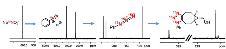
Using 15N-SABRE to probe a reaction pathway. Signals associated with each key species were detected in the spectrum.
Professor Duckett and his team then went on to demonstrate that they could detect the signals of these 15N species, in a test tube, in a magnetic resonance imaging experiment – the first step in translating the method towards MRI. As can be seen from the Figure, it was possible to achieve high resolution imaging.
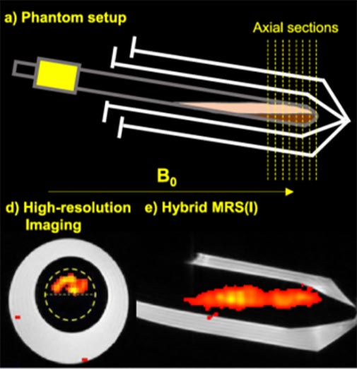
High resolution imaging achieved using 15N-SABRE.
Professor Duckett said: “Taken together, the results of this work indicate that it is possible to use SABRE to achieve high intensity, long-lived magnetic resonance signals which can be used to follow 15N-labelled reaction process. In the future, we would hope to develop the approach further to follow the distribution and metabolism of hyperpharmaceuticals within a living organism, potentially allowing MRI to achieve transformative steps forward in terms of diagnostic imaging.”
This research has been published in Journal of the American Chemical Society