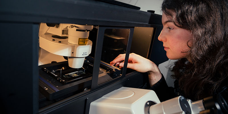
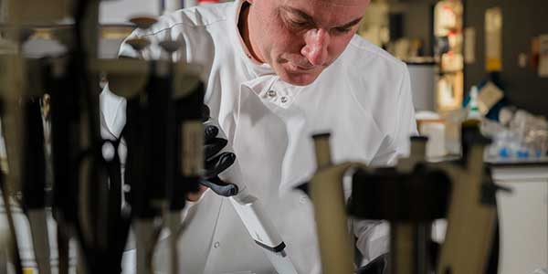
UK Biological Imaging Facilities

To search this site use Ctrl+F and enter term


View UK Biological Imaging Facilities in a larger map including Facility listing

Please scroll down the table (alphabetical order) or click the map link for more information about each facility

To add your facility to the list please contact Jo Marrison: jlm2@york.ac.uk


Institution Equipment

Aberdeen

Kevin Mackenzie
+44 (0)1224 437447

Miep Helfrich
+44 (0)1224 437443

Microscopy and Cellular Imaging Facility
Histology and EM Facility
Institute of Medical Sciences
University of Aberdeen
Foresterhill
Aberdeen
AB25 2Z

Access

  • Internal on all instruments
  • External commercial & academic on application
  • DIY or Full service work

Expertise
Core Facility; Histology, Electron Microscopy, MicroCT

Confocal microscopy
Zeiss 700 Laser Scanning Confocal Microscope
Zeiss 710 Laser Scanning Confocal Microscope
Zeiss 510 META Laser Scanning Confocal Microscope
Fluorescence microscopy
Zeiss Imager M2 + Apotome
Zeiss Observer Z1
Zeiss PALM Laser Microdissection system
Calcium imaging
Improvision system for time-lapse imaging
Micro CT
SkyScan-1072 High-resolution desk-top micro-CT system
Live cell Imaging
Delta Vision Core wide field imaging system with EMCCD
Histology
Tissue processors, standard and motorized microtomes for wax and resin sectioning
Cryostat for frozen sectioning
Leica Bond-max fully automated immuno-stainer
Electron Microscopy
2x Philips CM10 TEMs with Gatan CCD digital imaging
Zeiss EVO MA10 SEM with Oxford Inca microanalysis
Leica UC6, Ultracut S & Ultracut E ultramicrotomes
Cryo Electron Microscopy
Leica Empact 2 High Pressure Freezer (with rapid transfer system)
Leica AFS (Automated Freeze Substitution) processor
Leica FC6 cryo-ultramicrotome

Belfast

Stewart Church
+44 (0)2890972274

Bioimaging Core Technology Unit
School of Medicine Dentistry and Biomedical Sciences
Queens University of Belfast
Medical Biology Centre
97 Lisburn Road
Belfast
BT9 7AB
Northern Ireland

Access

  • Internal on all instruments
  • External commercial & academic on application
  • DIY or full support service
  • Proof of principle/ developmental studies undertaken

Expertise
Core Facility, dedicated staff for each imaging platform. Computational methods for quantitative image analysis. Content Management Systems for all images, central server with remote access.
Workshops, training courses, teaching. Embedded Cat 2 facilities with microbiological safety cabinets and animal holding suite.

Confocal Microscopy
Leica TCS SP2 with DMRBE/DMIRE2
Leica TCS SP5 with DMI6000+Solent live cell chamber (Cat 2 facility)
Fluorescence Microscopy
Zeiss Axiovert 200 with Axiovision
Leica DMI6000B with FW4000
Live Cell Imaging
Zeiss Axiovert 200 with Eppendorf Injectman +Femtojet (Cat 2 facility)
Virtual Microscopy
Aperio CS Scanner
Aperio OS Scanner
Hamamatsu Nanozoomer
in vivo Imaging
Xenogen IVIS 200
FACS
BD FACSAria II (Cat 2 facility)

Birmingham, BALM

Manuel Forero (Imaging Specialist)
+44 (0)121 414 3392

Alicia Hidalgo
(co-director of BALM)
+44 (0)121 414 5416

Birmingham Advanced Light Microscopy Facility (BALM)
School of Biosciences
University of Birmingham
Edgbaston
Birmingham
B15 2TT

Access

  • Internal, mostly used by School of Biosciences
  • External academic: used by many other Schools within this University
  • External commercial subject to access charges
  • Can be used as DIY or with technical help

Expertise
Core facility, confocal microscopy.
Fixed, time-lapse and cell culture.
Computational methods for quantitative image analysis, object recognition and cell tracking.

Future requirements
Computational methods for imaging

Confocal microscopy
Leica SP2 AOBS inverted (4 lasers)
BioRad Radiance 2000 upright (3 lasers)

Birmingham, BALM-AWN

Joshua Rappoport

(co-director of BALM)
+44 (0)121 414 9019

Birmingham Advanced Light Microscopy - Advantage West Midlands Imaging Facility (BALM-AWM)
School of Biosciences
University of Birmingham
Edgbaston
Birmingham
B15 2TT

Access

  • Internal (university-wide)
  • External

Expertise
Core facility - Live cell imaging; Probe development and high resolution imaging

Confocal microscopy
Nikon A1R confocal/TIRF with spectral detector, EM-CCD, and Piezo-Z drive

Bristol

Mark Jepson (light microscopy)
+44 (0)117 33 12143

Paul Verkade (electron microscopy)
+44 (0)117 33 12179

Prof. Paul Martin
+44 (0)117 3312298

Wolfson Bioimaging Facility
School of Medical Sciences
University of Bristol
University Walk
Bristol
BS8 1TD

Access

  • Currently most use is Bristol-based and subject to access charges
  • Scope for external collaborations in speciality areas
  • We work on a recharge basis both for full service (EM) as well as DIY.

Expertise
Core facility.
CLEM - Correlative light and electron microscopy. Fully integrated Light and Electron Microscopy Unit. Live cell imaging (especially intracellular trafficking, model organisms, bacterial infection).

Future requirements
Tools for image analysis, especially tracking.

Confocal microscopy
4 Leica CLSM systems: inverted SP5 AOBS, SP2 AOBS, SP2; upright NT
Spinning disk confocal: Perkin Elmer UltraVIEW ERS 6FE
Wide-field microscopy
3 Leica AS-MDW
2 Leica microscopes with Hamamatsu cameras, automated filter wheels and shutters, Improvision Volocity software
TIRF (Leica AM TIRF MC system for total internal reflection fluorescence microscopy or wide-field imaging)
Electron microscopy
FEI Tecnai 12: 120kV transmission electron microscope
FEI Tecnai 20: 200 kV transmission electron microscope equipped with a STEM unit; suitable for cryo-EM and electron tomography, including STEM tomography.
FEI Quanta 400 variable pressure scanning electron microscope
Extensive EM preparation facilities.
Other
Offline image analysis suite for LM and EM.
Almost all Light Microscopes are equipped with live cell imaging chambers.
Cell culture facilities within the facility.

Cambridge

Richard Adams
+44 (0)1223 333782

Department of Physiology, Development and Neuroscience
University of Cambridge
Downing Street
Cambridge
CB2 3DY

Expertise
Time-lapse imaging, image processing, computational methods

Image analysis.
Cell tracking and cell shape analysis in whole embryos.
Computational analysis of tissue dynamics during embryogenesis.

Cambridge

Matt Wayland
+44 (0)1223 769282

Imaging Facility
Department of Zoology
University of Cambridge
Downing Street
Cambridge
CB2 3EJ

Access

  • Internal (80% on all instruments)
  • External commercial & external academic (20% on all)
  • DIY or full service work

Expertise
Core Facility
Image analysis and programming

Confocal microscopy
Leica SP5 invert
Leica SP5 upright
Leica SP upright
UltraView CSU-10 Spinning Disk
MicroPoint laser ablation / microdissection
Live cell imaging chamber on SP5 invert (up to 37oC and 5% CO2

Cambridge

Alexandre Kabla
+44 (0)1223 766208

Department of Engineering
University of Cambridge
Trumpington Street
Cambridge
CB2 1PZ

Expertise
Imaging, computational methods

Image Analysis
Image processing and mechanical properties of multicellular assemblies

Cambridge

Clemens Kaminski
+44 (0)1223 763135

Laser Analytics Group
Department of Chemical Engineering and Biotechnology
University of Cambridge
Pembroke Street
Cambridge
CB2 3RA

Access

  • Internal
  • External
  • DIY

Expertise
All aspects of quantitative multidimensional microscopy

Confocal microscopy
Custom built widefield FLIM platform with LED and Supercontinuum excitation (430-700 nm). Both ultrahigh speed FLIM (15 fps at full field) and excitation at multiple modulation frequencies are possible.
Custom designed hyperspectral imaging confocal station, with supercontinuum excitation (430-700 nm excitation / emission, at electronically selectable, arbitrary wavelength combinations). Full resolution of fluoresence excitation and emission spectrum, fluorescence lifetime and polarisation state. TCSPC, Fluorescence Anisotropy Imaging
Leica SP5 confocal platform with resonant scanner.

Cambridge

David Klenerman
+44 (0)1223 336481

The University Chemical Laboratory

Dept of Chemistry
University of Cambridge
Lensfield Road
Cambridge
CB2 1EW

Expertise
Laser fluorescence spectroscopy and scanning probe microscopy

Fluorescence microscopy
Two colour total internal reflection single molecule fluorescence microscope

Cambridge

Jeremy Skepper
+44 (0)1223 333774

Janet Powell
+44 (0)1223 333774

Multi-Imaging Centre
Anatomy Building
Physiology Development and Neuroscience,
University of Cambridge
Downing Site
Cambridge
CB2 3DY

Access

  • Internal
  • External
  • Commercial.
  • DIY and service work.

Expertise
Specimen preparation, ambient and low temperature, stereology, x-ray microanalysis, immunocytochemistry, bio-materials microscopy, physiological and time-lapse microscopy.

Confocal microscopy and LMD
Leica SP1- multiphoton (invert)
Leica SP1-UV (invert)
Leica SP2-405 - 633 (invert)
Leica SP2-456 - 633 (invert)
Leica SP5-456 - 633 (invert physiology)
Leica LMD 6000 (upright laser microdissection microscope)
Electron microscopy
FEI Technai G2 200 kv, STEM, tomgraphy, cryo-transfer, HAADF, SAED, CBED, EDAX Genesis EDX, Deben AMT digital camera
FEI-Philips XL30 FEGSEM, SE and BSE detectors
Oxford INCA EDX with 30mm pentafet ATW detector
Quorum Emitech PP2000 cryotransfer system
Specimen preparation
Leica VT1000S vibrating blade microtome
Leica CM 3050S Cryostat
Leica CPC plunge and impact freezing device
2 x Leica AFS freeze substitution and low temperature embedding devices
3 x Leica Ultracut E
Leica Ultracut UCT
Leica Ultracut S and FCS cryosectioning device
Edwards 306 auto, Carbon coater, freeze drier and glow discharge unit
Quorum Emitech K575X dual head Au and Cr sputter coater and remote pumping station for TEM cryo-transfer specimen holder
Polaron CPD

Cambridge, Babraham

Simon Walker
+44(0)1223 496618

Imaging Facility
The Babraham Institute
Babraham
Cambridge
CB22 3AT

Access

  • Full access to external users, though internal users get priority.
  • DIY or Full service work

Expertise
Core Facility
Live cell fluorescence imaging

Confocal microscopy
Zeiss LSM 510 META invert
Olympus FV1000 invert (x2)
Spinning Disk with EM CCD (Andor)
Fluorescence imaging
Olympus CellR (widefield+TIRF)
Live cell imaging chamber
High Content Screening
BD Pathway 855
MetaSystems Metafer / MetaCyte

Cambridge, Centre for Stem Cell Research

Peter Humphreys (light microscopy)
+44(0)1223 760200

Rachael Walker (flow cytometry)
+44(0)1223 760227

Centre For Stem Cell Research
Tennis court Road
Cambridge
CB2 1QR

Expertise
Core Facility, image analysis

Future requirements
HE/fluorescent upright
Plate scanning microscope

Confocal Microscopy
Leica SP5 invert + live cell chamber x2
Andor Revolution XD spinning disk
Widefield and cell culture
Nikon Biostation IM
Essen Incucyte HD
Essen Incucyte
Zeiss AxioExaminer
Leica DMI4000
Olympus IX51 x3
Flow Cytometry
MoFlo
CyAN

Cambridge, Gurdon Inst.

Alex Sossick
+44 (0)1223 334082

Wellcome Trust/Cancer Research UK Gurdon Institute
University of Cambridge
Tennis Court Road
Cambridge
CB2 1QN

Access

  • Priority given to Gurdon Institute users
  • Limited outside access

Expertise
Core Facility. Immunofluorescence; widefield epifluorescence and confocal microscopy, deconvolution

Confocal microscopy
Confocal scanning laser microscopes and multi-photon system
Widefield deconvolution and high throughput systems

Cardiff

Jim Ralphs
+44 (0)2920 874954

Tony Hayes
+44 (0)2920 876611

Marc Isaacs
+44 (0)2920 876611

Confocal Microscopy Unit
Cardiff School of Biosciences
Cardiff University
Cardiff
CF10 3AX

Access

  • Internal (mainly), but also external commercial & academic

Expertise
Core Facility; Immunofluorescence; Widefield epifluorescence and confocal microscopy

Confocal microscopy
Leica TCS SP2 AOBS laser scanning confocal microscope. Twin microscope configuration comprising upright (DM6000) and inverted (DMIRE2) stands (laser lines: 405, 458, 476, 488, 514, 543 & 633nm), galvonometer & electromotor stages; Solent Scientific incubation chamber with CO2/humidity and temperature regulators for in vitro work; brightfield, fluorescence, DIC and polarizing optics
Fluorescence imaging
Olympus BX series microscopes with fluorescence, DIC and brightfield optics refer to web pages for further information

Cardiff

Anna Hurley
+44 (0)2920 687004

Charlotte James
+44 (0)2920 687004

Central Biotechnology Services
Henry Wellcome Building
School Of Medicine
Cardiff University
Heath Park
Cardiff
CF14 4XN

Access

  • Internal and external users
  • Academica and Industry
  • User access or managed service

Expertise
Core facility, academic consultancy, certified to ISO 9001 and ISO 17025

Confocal microscopy
Leica SP5
Leica SP2
Flow cytometry
3 x FACS Calibur 4 colour
1 x Beckman Cyan ADP 9 colour
1 x FACS Canto II 8 colour
1 x FACS Calibur 3 colour
Beckman MoFlo - 9 parameters simultaneously using 6 colours

Cardiff

Jan Hobot
+44 (0) 2920 742131

Chris von Ruhland
+44 (0) 2920 742132

Medical Microscopy Sciences
School of Medicine
Cardiff University
Heath Park
Cardiff
CF14 4XN

Access

  • Internal and external both academic and commercial.

Expertise
Core facility.
Specimen preparation at ambient or low temperature for structural or immunocytochemical studies.
First-hand knowledge of modern acrylics (Lowicryls and LR White).
Tokuyasu or cryosubstitution methodologies.
Histology and LM preparations.
TEM and SEM X-ray microanalysis.
UKAS accreditation for X-ray microanalysis of materials and particulates for detecting asbestos.

Fluorescence imaging
Olympus BX51 Upright with Zeiss AxioCam camera.
Olympus CK40 Inverted with Polaroid digital camera.
Reichert Polarising Microscope.
Electron microscopy
Philips CM12 TEM with SIS MegaView III camera and EDAX Genesis X-ray microananalysis system.
JEOL 840A SEM with ADDA SIS digital imaging and INCA x-sight detector X-ray microanalysis system with elemental mapping plus Oxford CT1500 Cryosystem and stage.
Specimen Preparation
Leica EMTP Tissue Processor.
Reichert KF80 metal mirror/plunge freezer.
Dedicated Histology room.

Didcot, STFC

David Clarke
+44 (0)1235 567035

Octopus Imaging Cluster (Lasers for Science Facility)
Science & Technology Facilities Council
Rutherford Appleton Laboratory
Didcot
OX11 0QX

Access

  • Via peer-review panel for external and internal users
  • Approx 80% of scheduled time for long-term (>1 year) programmes, and 20% for proof-of-principle work
  • Additional access for users of other facilities (Diamond Light Source, ISIS, Research Complex at Harwell)
  • Commercial access also available

Expertise
Core Facility; Single molecule techniques, including in live cells; single molecule and ensemble FRET; FLIM; development of advanced laser microscopy instrumentation.

Future requirements
Image analysis for single molecule work

Central laser cluster
(ns, ps, fs and steady state lasers UV-near IR) feeding the following microscopy stations:
Single molecule microscopy
Multidimensional (simultaneous two-colour(FRET)/two polarisations) single molecule TIRF microscope (live cell and immobilised samples).
3-colour single molecule TIRF X-Y-t tracking (Multiprotein complex kinetics and dynamics).
5-colour single molecule TIRF X-Y-t tracking (Multiprotein complex kinetics and dynamics) - under construction - to be scheduled next year.
Confocal microscopy
Multiphoton confocal fluorescence microscope for FRET/FLIM Imaging.
Multicolour confocal fluorescence microscope with Becker & Hickl Fluorescence Lifetime Imaging (FLIM) module.
Support Equipment
Laser tweezers, steady-state and time-resolved laser spectroscopy apparatus, cell biology and biochemistry support labs

Dundee

Jason Swedlow
+44 (0)1382 385819

Swedlow lab
Wellcome Trust Centre for Gene Regulation & Expression
College of Life Sciences
MSI/WTB/JBC Complex
University of Dundee
Dow Street
Dundee
DD1 5EH

Open Microscopy Environment

Expertise
Structured illumination microscopy on OMX, expertise on high content screening and live cell imaging, development of data management tools and specifications for microscopy.

Future requirements
Money for meetings and travel to other sites for access and sharing of info and smart, programmable microscopes.


Dundee

Sam Swift
+44 (0)138 238 6412

Central Microscopy Facility
College of Life Sciences
MSI/WTB Complex
University of Dundee
Dow Street
Dundee
DD1 5EP

Access

  • User training/DIY for light microscopy
  • DIY or Full Service for EM
  • External academic / industry welcome by appointment

Expertise
Core facility; High resolution imaging, live cell imaging, FLIM, FRET, FRAP, immuno EM etc

Light Microscopy
BioRad Radicance 2100MP confocal / multiphoton
Zeiss LSM 510 META
Leica SP2
Becker and Hickl TCSPC FLIM
DeltaVision Personal, RT and Core restoration microscopes with EMCCD, Live Cell Chambers and Laser Modules
Nikon TIRF
OMX Structured Illumination Microscope
Electron Microscopy
FEI Tecnai 12 TEM
Jeol 1200 EX TEM
Hitachi s4700 FESEM

Dundee

Markus Posch (SULSA Scientific Officer for OMX)
+44 (0)138 238 6414

OMX Super-Resolution Microscopy
College of Life Sciences
University of Dundee
Dow Street
Dundee
DD1 5EH

Access

  • Any users welcome
  • Euro BioImaging Proof of Concept site(Advanced Light Microscopy - General Access)

Expertise
Structured Illumination Spatial Super-Resolution - Superfast Live Imaging

OMX ver. 3 platform (Applied Precision)

Durham

Tim Hawkins (Advance Light Microscopy)
+44 (0)191 334 1333

Christine Richardson (Electron Microscopy)
+44 (0)191 334 1285

Microscopy and Bioimaging Facility
School of Biological & Biomedical Sciences
Durham University
Durham
DH1 3LE

Access

  • Internal on all instruments
  • External commercial & external academic
  • DIY or Full Service

Expertise

Core facility; High resolution imaging, FRET, FRAP, FLIP. 5D live cell imaging, TIRF, photoactivation/conversion,
ImmunoGold, Cryosectioning, Ultramicrotomy, Sterology.
Licenced to image plant:pathogen interactions within facility.
Collaboration & developments of new techniques (eg photonics, chemical probes)

Future requirements
SuperResolution, FCCS & FLIM
Confocal microscopy
Leica TCS SP5 Tandem head AOBS Invert (Resonant scanner)
Zeiss LSM 510 META invert
BioRad Microradiance Upright
Andor Revolution XD Spinning Disk Invert
Fluorescence Imaging
Leica MC TIRF Microscope
Zeiss Apotome Microscope
Zeiss Live Cell Imaging System
EM CCD Imaging
Most of the above microscopes are within live cell imaging/environmental chambers
Microinjection
Upright (DM2500) and inverted (DMI3000) DIC microscopes + multiple injection systems to work with a wide variety of organisms and tissues. HD imaging system.
Electron Microscopy
Hitachi H7600 TEM
High resolution Hitachi S5200 FESEM + Cryostage
Image analysis
Suite of high-end 64-bit PC workstations
Deconvolution
Sample Preparation
Leica High Pressure Freezer EMPACT
Leica AFS freeze substitution and low temperature
Leica Ultracut S room temperature
Leica FC6 cryo-ultramicrotome
Leica Knife maker
Baltec Critical Point Drier
Cressington coating units (Carbon, platinum, chromium, tungsten)
Leica CPC cryo workstation
Leica IGL. Automatic immunogold labeller
Leica MM80E metal mirror/plunge freezer
Tissue processing
Cryostat for frozen sectioning
Microtomes for wax and resin sectioning
Leica vibrating diamond knife

Edinburgh

Rolly Wiegand (LM)
+44 (0)131 650 2864
CALM - Confocal and Advanced Light Microscopy Facility

Shonna Johnston (Flow)
+44 (0)131 242 6567

Flow Cytometry Facility

Queen's Medical Research Institute
47 Little France Crescent
Edinburgh
EH16 4TJ

Access

LM

  • Internal for University
  • External
  • DIY or fully assisted image acquisition

Flow

  • Internal for University only
  • Fully assisted cell sorting

Expertise
Core Facility; Teaching - Microscopy course (twice a year), high resolution cellular microscopy, image restoration and quantitation, live cell imaging

Future requirements
Hardware for deep penetration in/ex vivo live cell imaging and TIRFM

Confocal microscopy
Zeiss LSM 510 META, inverted
Leica SP5, inverted, both eqipped with environmental control
Fluorescence imaging
Olympus Cell P, inverted
Environmental control for live cell imaging
Optical Projection Tomography (Bioptonics 3001)
Image analysis
High-end workstations with SVI Huygens Essential, ImageJ and PE Volocity

Flow cytometry
Cytometry analysers
BD FACSCalibur
BD FACScan
BD FACSArray
Cell sorters
BD FACSVantage SE (Diva)
BD LSR Fortessa
BD FACSAria II

Edinburgh, Clinical Research Imaging Centre (CRIC)

Edwin J. R. van Beek
+44 (0)131 242 7760
SYNAPSE Chair of Clinical Radiology

Stuart Docherty
+44 (0)131 242 7767

Clinical Research Imaging Centre

University of Edinburgh
CO.19
Queen's Medical Research Institute
47 Little France Crescent
Edinburgh
EH16 4TJ

Access

  • Access through applications process
  • PET-CT 70% NHS service
  • MDCT 50% NHS service
  • MRI 100% research


Expertise
Core facility.
Imaging-based assessment of structure, function and treatment efficacy

Imaging
3T MRI (Siemens Verio) with fMRI, multinuclear spectroscopy and MR Elastography
Mock MRI system
320 MDCT (Toshiba Aquilion ONE)
128 mCT-PET (Siemens)
PET-trace 8 cyclotron (GE)
Radiochemistry
4 GMP hot cells and 3 research hot cells

Image analysis laboratory

Edinburgh, Orthopaedic Engineering Collaboration

Antonello Spadaccino
+44 (0)131 242 6295

Orthopaedic Engineering Collaboration
Chancellor's Building
University of Edinburgh
Room FU 413
49 Little France Crescent
Edinburgh
EH16 4SB

Access

  • Internal (75%)
  • External and commercial (25%)
  • Full service work
  • Induction training and supported DIY

Expertise
Scan and production of 3D images from samples.
Comprehensive tensile, 3 & 4 point bend tests.
Torsional testing, ultimate strength, fatigue and step loading. Combination of axial and torsional tests.


Xray Imaging
Skyscan 1172
Mechanical Testing
Zwick/Roel z0005


Edinburgh, IMPACT

Trudi Gillespie
+44 (0)131 6503278

IMPACT Imaging Facility
Hugh Robson Building
George Square
Edinburgh
EH8 9XD

Access

  • Internal for University
  • External
  • DIY or fully assisted image acquisition

Expertise
Core Facility.
High resolution cellular microscopy, image restoration and quantitation, live cell imaging, TRIF and FLIM

Future requirements
MP system for in/ex vivo live cell imaging

Confocal microscopy
2 Zeiss LSM 510 one inverted and one upright both coupled to a Coherent Mira mode-locked Ti:Sapphire multi-photon laser equipped with environmental control and a Becker and Hickl unit for fluorescence lifetime imaging (FLIM).
Leica TCS-NT, inverted and upright with environmental control
Fluorescence imaging
Olympus Cell R Dual TIRF System, with environmental control for live cell imaging
Image analysis
High-end workstations with SVI Huygens Essential and Pro, ImageJ, Imaris and PE Volocity

Edinburgh, MRC

Mike Millar
+44 (0)131 242 9155

Histology and Imaging Facility
MRC, Human Reproductive Sciences Unit
The Queens Medical Research Institute
New RIE
47 Little France Cres
Edinburgh
EH16 4TJ

Access

  • Internal on all instruments
  • External academic on application
  • DIY or Full service work

Expertise
Core facility.
Confocal microscopy, Stereology and immunohistology with emphasis on multi same species colocalisations.
Reproductive morphology

Confocal microscopy
Zeiss 510 Laser Scanning Confocal Microscope
Zeiss 510 META Laser Scanning Confocal Microscope
Zeiss 710 Laser Scanning Confocal Microscope
Widefield microscopy
Assorted upright and invert research grade microscopes with CCD imaging both in colour and monochrome
Live cell Imaging
Stereology using Media Cybernetics Image Pro Plus with stereologer plug in
Image Analysis
Huygens deconvolution software
Volocity 3D imaging software
Media Cybernetics Image Pro Plus
Histology
Tissue processors, standard and motorized microtomes for wax and resin sectioning
Cryostat for frozen sectioning
Leica Bond- fully automated immuno-stainer
Intavis whole mount in-situ hybridisation and immunostaining robot

Edinburgh, Brain Research Imaging Centre (BRIC)

Joanna Wardlaw (Director of Centre)
+44 (0)131 537 2943 (PA)

Duncan Martin (Scientific Business Manager)
+44 (0)131 537 2664

Brain Research Imaging Centre
University of Edinburgh
Division of Clinical Neurosciences
Bramwell Dott Building
Western General Hospital
Edinburgh
EH4 2XU

Access

  • Access through an application process
  • Research and commercial scanning work can be undertaken

Expertise
Stroke, FMRI, Diffusion and tractography, Perfusion imaging, Aging, Spectroscopy, Psychiatric disorders, Neurodegenerative disorders
1.5T (GE) MRI imaging capabilities
Structural
Diffusion and Tractography
Functional MRI
(Simultaneous EEG)
Perfusion and Bloodflow
Permeability
Spectroscopy
Temperature Mapping

Radiological advice on imaging and study protocols
Radiological interpretation of scans

Image Analysis methods

MRI Quality Assurance methods

Software tools for image analysis and image management

Essex

Philippe Laissue
+44 (0)1206 872246

Bioimaging Facility
Department of Biological Sciences
Wivenhoe Park
University of Essex
Colchester
CO4 3SQ

Access

  • Internal on all instruments
  • External commercial & academic on application; Nikon reference site
  • Proof of principle studies undertaken

Expertise
Core facility, rapid and long term live cell imaging, immunofluorescence, colocalisation, visualisation. Development of specialised instruments and computational methods for quantitative image analysis.

Future requirements
Further in-house development of instruments and algorithms. Super-resolution and FLIM.

Confocal microscopy
Nikon A1R (inverted) with 6 laser lines, hybrid scanner, spectral detector, automated XY and piezo Z,
TIRF/OAF/widefield with EM-CCD, live cell chamber
Bio-Rad Radiance 2000 with Olympus IX70 (inverted)
Widefield fluorescence Microscopy
Nikon Ti-E (inverted) with LEDs and Intensilight, EM-CCD, automated XY and piezo Z, live cell chamber
In-house developed TIRF and OAF instrument (inverted) with EM-CCD and triple split (Kad group)
Various upright and inverted fluorescence and brightfield microscopes with EM-CCD and colour CCD cameras
Several commercial and in-house developed imaging systems for measurement of photosynthesis by gas exchange and fluorescence, both in the laboratory and in the field (Lawson group).
Image Analysis
High end 64-bit workstations for offline image processing and analysis. NIS-Elements, deconvolution. Custom software using ImageJ and Matlab. Workshops, training courses, teaching. Extensive website for booking, training, resources.
Other
Fully equiped Cat 2 laboratory, cell culture facilities

Flow Cytometry
BD FACS Aria
BD FACS Calibur


Glasgow, Beatson

Kurt Anderson
+44 (0)141 330 2864

Beatson Institute for Cancer Research
Garscube Estate
Switchback Road
Glasgow
G61 1BD

Access

  • Priority given to Beatson users, followed by Glasgow University
  • Outside access primarily on a collaborative basis

Expertise
Live cell imaging, including time and frequency domain FLIM, TIRF, photo-activation / -bleaching / -conversion.
Mouse in vivo imaging, including collagen SHG and use of photo-activation and -bleaching, FLIM and FRET.

Future requirements
Image analysis, adaptive optics

Confocal microscopy
Olympus FV1000 (x2)
Nikon A1R
Leica TCS SP2
Zeiss LSM 710

FLIM
LaVision Bio-Tec TRIM scope
Lambert Instruments custom Spinning Disk
Lambert Instruments custom TIRF
Leica TCS SP2 with Becker & Hickle TCSPS 730

Live Cell
Custom TIRF
Olympus / Volocity Deconvolution system
Micro-injection
Medium Throughput Long Term Time Lapse (x4)
 
Other
Olympus OV100
Leica AF6000 micro-dissection
FacsAria
Upright stands for histology and immunofluorescence (x4)
Off-line image analysis (x8)

Leeds

Gareth Howell
+44 (0)113 3437270

Leeds Bioimaging and Flow Cytometry Facility
Faculty of Biological Sciences
University of Leeds
Leeds
LS2 9JT

Access

  • Internal and external, academic research groups and commercial

Expertise
Core Facility.
Collaboration and development of techniques, image processing and analysis. Live cell imaging.

Confocal microscopy
Zeiss LSM 510 META invert with motorised stage and 405 laser diode
Zeiss LSM 510 META upright with motorised stage
Fluorescence imaging
Delta Vision Deconvolution System
Live cell imaging chambers
Microinjection
Eppendorff FemtoJet system on an inverted Zeiss Axiovert 200
Flow cytometry
FACSCalibur 4 colour analyser flow cytometer
FACSAria 7 colour cell sorter
Partec PASIII 4 colour flow cytometer

Leicester

Kees Straatman
+ 44 (0)116 229 7085

Advanced Imaging Facilities
Department of Biochemistry
Henry Wellcome Building
University of Leicester
Lancaster Road
Leicester
LE1 9HN

Access

  • Mostly internal users but external users are welcome

Expertise
Core Facility.
Immunofluorescence, widefield epifluorescence, confocal microscopy, live cell imaging, microinjection, FRAP/FLIP/FRET, spectral imaging, high content screening/analysis.
Data analysis and quantification: Imaris, Volocity, Openlab, Huygens Essential deconvolution, ScanR/CellR, ImageJ, NIS Elements.
Teaching: Microscopy workshops, lectures

Future requirements
Technical support
Image indexing software
FCS/FLIM
Super resolution imaging
Whole animal imaging (funding secured)

Most systems with incubators, temp and CO2 control
Confocal microscopy
Leica TCS SP5 inverted
Olympus FV1000 inverted
Nikon C1Si inverted
Zeiss LSM 7 MP upright (soon)
Fluorescence imaging
3 Nikon TE 300 inverted
1 Nikon Ti-E EM-CCD + LEDs inverted
Olympus CellR/scanR inverted
1 Nikon TIRF EM-CCD inverted
Olympus BX61 upright with Applied Spectral Imaging's scanning platform
Bioluminescence imaging
Olympus LV200 EM-CCD
Flow cytometry
BD FACS Canto II
Imagers
Typhoon Trio+
LAS4000

London, Imperial

Raffaella Carzaniga
+44 (0)207 594 5194

Electron Microscopy Centre
Imperial College
Exhibition Road
South Kensington
Biochemistry Building- Room 022
London
SW7 2AZ

Access

  • Internal and external academic research groups and commercial

Expertise
Cryo-Preparation methods for TEM, TEM for Cellular Biology & Single Particles.
Image processing for Single Particles

Electron microscopy
TEM-Philips CM100 (Tungsten electron source) with film plate
TEM- FEI Tecnai 12 (LaB6 electron source); CCD camera
TEM Philips CM200 (FEG electron source); CCD camera
TEM- Philips CM300 (FEG electron source); Helium cooled-Polara Stage; CCD camera
Vitrobot
HPF- High Pressure Freezing apparatus (EMPACT 2 with RT, Leica)
Ultramicrotomes (for ambient and low temperatures)
Parallel computer farm

London, Imperial

Dirk Dormann
+44 (0)208 383 8528

Microscopy Facility
MRC Clinical Sciences Centre
Faculty of Medicine Imperial College London
Hammersmith Hospital Campus
Du Cane Road
London
W12 0NN

Access

  • Core funded by MRC, only MRC users

Expertise
Core facility.
Quantitative image analysis, time-lapse

Confocal Microscopy
Leica SP1 invert
Leica SP2 invert
Leica SP5 invert with tandem scanner and FCS/FCCS
PerkinElmer UltraView Spinning Disk
Fluorescence imaging
Deltavision Spectris
Leica DMIRE2 inverted with Eppendorf microinjection system
Leica DMRB upright
Zeiss Axiovert 100S TV inverted
Zeiss Axiophot upright
Olympus SMZ-16X stereo microscope
PALM laser capture/microdissection

London, Imperial

Paul French
+44 (0)207 594 7713

Photonics Group
Physics Department
Imperial College London
South Kensington Campus
London
SW7 2AZ

Access

  • By collaboration

Expertise
Optical Microscopy, Optical Tomography, Optical Endoscopy, Imaging Data Management and Integration, Optical Image Analysis

Future requirements
Multidisciplinary training at the interface of photonics technology and life science applications.
Long term funding for core instrumentation staff.
Tb scale optical image data management systems.
Software tools including automatic image segmentation and multidimensional analysis.

Multidimensional fluorometer
Confocal/Multiphoton Microscope, Ti-Sapphire laser or supercontinuum excitation source/FLIM/polarisation-resolved imaging
Line-scanning semiconfocal microscope with Ti-Sapphire laser or supercontinuum excitation source
TIRF microscope, multispectral imaging, FLIM/polarisation resolved imaging capability
Wide-field FLIM microscope optimised for UV excitation
Multibeam multiphoton microscope (LaVision TriMScope) with FLIM
High throughput automated optically sectioned (Nipkow) FLIM microscope
High speed Nipkow disc FLIM microscope with supercontinuum excitation
STED FLIM microscope, Leica SP2 laser scanning confocal, Ti:sapphire laser
Wide-field FLIM endoscope/macroscope system
Microconfocal FLIM endoscope
FLIM Optical Projection Tomography (OPT)

London, Imperial

Martin Spitaler
+44(0)207 594 2023

FILM (Facility for Imaging by Light Microscopy)
Imperial College Exhibition Road
South Kensington / SAFB
London
SW7 2AZ

Access

  • Internal and external (academic and commercial)
  • DIY or assisted microscopy
  • Assay development

Expertise
Core facility

Confocal microscopy
Zeiss LSM-510
Leica SP2
Leica SP5 resonant
Leica SP5 MP-Flim upright and inverted
Widefield microscopy
EM-CCD
Incubation chambers
Automated XYZT imaging
Low and medium throughput
TIRF
Image analysis

Volocity
SimplePCI
Autoquant
CellProfiler
Matlab
Definiens

London, King's

Rainer Heintzmann

Matthias Gautel

Randall Division of Cell and Molecular Biophysics
King's College London
New Hunt's House
Guy's Campus
London
SE1 1UL

Access

  • Internal
  • Collaborations to apply superresolution imaging are possible
  • DIY

Expertise
Superresolution Microscopy.
Facilities for structured illumination, 4pi, STED (soon), expertise in deep tissue imaging, FRET and FLIM.

Future requirements
Financial support for personnel for applying existing expertise to critical biological problems

Structured Illumination
Lecia 4Pi TCS SP2
Leica STED

London, King's

Christopher Healy
+44 (0)207 188 1795

Tissue Imaging Centre,
Dept of Craniofacial Development,
King's College London Dental Institute at
Guy's,
King's College and St Thomas' Hospitals,
Floor 27 Tower Wing,
Guy's Hospital,
St. Thomas St.,
LONDON
SE1 9RT

Access

  • Internal (100% on microscopes, 90% on microCT)
  • External commercial & external academic (10% on all)
  • DIY on microscopes.
  • Part or Full service work on MicroCT and OPT

Expertise
Core facility.
Morphometrics, bone histomorphometry and high resolution imaging.

Confocal microscopy
Leica SP5 upright confocal microscope
Fluorescence imaging
Zeiss Axioskops with Axiocamera
PALM laser capture/microdissection
Live cell imaging chambers
microCT
GE Healthcare Locus SP specimen microCT
Optical Projection Tomography
Bioptonics 3001

London, CRUK-LRI

Daniel Zicha (LM)
+44 (0)207 269 3563

Lucy Collinson (EM)
+44 (0)207 269 3416

Anne Vaahtokari (Super-resolution microscopy)
+44 (0)207 269 3511

Cancer Research UK, London Research Institute
Lincoln's Inn Fields Laboratories
44 Lincoln's Inn Fields
London
WC2A 3PX

Access

  • LM - Priority given to LRI users, limited outside access
  • EM - Core Technology Facility, internal groups only
  • Super resolution - Priority given to LRI users, limited outside access

Expertise
Core facility.
Expertise in image processing and simulation.
Quantitative analysis with multi-modal microscopy.
EM - Preparation of cell biological material for TEM and SEM.
Cryo preparation techniques for biological samples (HPF, FS, Tokuyasu, freeze fracture, freeze drying, cryoEM)
Tomography
Correlative Light/ Electron Microscopy (CLEM)
Volume imaging (FIB/SEM, SBF/SEM, MicroCT, NanoCT)
Super-resolution microscopy

Future requirements
Support for developing new image processing approaches

Confocal microscopy
LSM 510 (Zeiss) including a multiphoton system
Leica SP5
Low light level imaging systems
Universal Imaging Metamorph,
Kinetic Imaging AQM
Molecular Devices Discovery-1
PerkinElmer UltraVIEW confocal imaging systems
Eppendorf Microinjection systems

Electron Microscopy Unit
Electron microscopy
FEI Tecnai G2 Sprit TEM 120kV, with tomography stage, single and dual tilt tomography holders, bottom mount Gatan Ultrascan CCD, side mount Gatan Orius CCD
FEI Tecnai G2 Sprit TEM 120kV, with cryo stage and cryo holder, bottom mount Gatan Ultrascan CCD, side mount Gatan Orius CCD
Jeol 1010 TEM with side mount Gatan 1kx1k CCD
Jeol JSM 6700F Field Emission SEM
Specimen preparation
Leica High Pressure Freezer EMPACT2 and RTS
2 Leica AFS freeze substitution and low temperature embedding devices (1 AFS1, 1 AFS2 with FSP)
2 Leica Ultracut UCT room temperature
Reichert Ultracut S room temperature
2 Leica Ultracut UCT, FCS cryo
Knife maker
Baltec Med020 dual gun sputter coater (carbon and carbon/platinum) and rotary shadower
2 Critical Point Drier 3000
Polaron sputter coater
Gatan ion beam coater
Emitech freeze substitution K775X
FEI Vitrobot Mark IV
Software expertise
Gatan Digital Micrograph
Inspect 3D
Amira
Imaris
Drishti

Super-resolution Microscopy
API OMX V3 structured illumination super-resolution microscope

London, CRUK-LRI

Francois Lassailly
+44 (0)207 269 3426

In vivo Imaging Facility
London Research Institute
Cancer Research UK
44 Lincoln's Inn Fields
London
WC2A 3PX

Access

  • Primarily for LRI users but could be discussed for external users


Expertise
Non-invasive in vivo imaging.
2D, 3D, 4D imaging.
Longitudinal studies for orthotopic models in oncology and hematology, normal development, stem cells, microenvironment, vascular and bone biology.
Assessment of validation of probes and contrasting procedures.
Image processing and analysis

Whole body Bioluminescence (including diffuse optical tomography and spectral unmixing)
Caliper Life Sciences IVIS-Spectrum
Whole body Fluorescence (including epifluorescence, transillumination, tomography and spectral unmixing from UV to IR))
Caliper Life Sciences IVIS-Spectrum
Micro-CT (including physiological monitoring for respiratory or cardiac gating, 4D)
Skyscan 1176 in vivo scanner
Intravital microscopy of live tissues (combining multiphoton and confocal, equiped with multiparametric physiological monitoring)
Zeiss LSM710-NLO


London, LSHTM

Theresa Ward
+44 (0)207 612 7869

Liz McCarthy and Rachel Gregory (LM)
+44 (0)207 927 2063

Maria McCrossan (EM)
+44 (0)207 927 2373

Liz King (Flow Cytometry)
+44 (0)207 927 2373

Woolfson Cell Biology Facility
Immunology Unit
London School of Hygiene & Tropical Medicine
Keppel Street
London
WC1E 7HT

Access

  • Internal (90% on all instruments)
  • External academic (10%)
  • DIY or Full service work

Expertise
Imaging pathogen-host interactions, at cellular level, and in whole model organisms. Includes live imaging under Cat 2 and full aerosol Cat3 containment.

Future requirements
Multiphoton for cell and in vivo imaging under containment

Confocal microscopy
Zeiss LSM 510 invert with live cell chamber and Eppendorf microinjeciton
Fluorescence imaging
Zeiss Axioplan 2 upright with CCD, epifluorescence and histology
Keyence BZ-8000 automated fluorescence microscope under Cat3 containment
Volocity software
Laser capture/microdissection
Arcturus Pixcell IIe
In vivo imaging under Cat3 containment
IVIS Spectrum (Xenogen)
Fluorescence molecular tomography (FMT) system
Electron microscopy
Jeol JEM-1200 EXII TEM
Jeol JSM-25S III SEM
Flow cytometry
2x Becton Dickinson FACSCalibur (one under Cat3 containment)
Becton Dickinson FACSVantage SE cell sorter

London, National Physical Laboratory

Alex Knight (Biotechnolgy Group)
+44 (0)208 9436308

Daniel Metcalf (Biotechnology Group)
+44 (0)208 9436344

National Physical Laboratory
Analytical Sciences Division
Hampton Road
Teddington
TW11 0LW

Access

  • Internal
  • External commercial contract research
  • External academic collaborations welcome

Expertise
Super-resolution microscopy, Single molecule fluorescence, Confocal microscopy, Adaptive optics, Optical coherence tomography, Instrumentation, Metrology/standards

Super-resolution Microscopy:
Direct stochastic optical reconstruction microscope (self-build)
Single molecule fluorescence microscope (self-build)
Structured illumination microscope (self-build, ready early 2012)
Photoactivated localisation microscope (self-build, ready early 2012)
Other imaging techniques available at NPL include:
Confocal fluorescence microscope (Olympus FV1000), Scanning ion conductance microscopy, atomic force microscopy, raman microscopy, secondary ion mass spectrometry, optical coherence tomography, X-ray photoelectron spectroscopy, matrix assisted laser desorption ionisation mass spectrometry, scanning electron microscopy, terahertz imaging, ultrasound


London, NIMR

Yan Gu
+44 (0)208 816 2621

Confocal Imaging and Analysis Lab
National Institute for Medical Research
The Ridgeway
Mill Hill
London
NW7 1AA

Access

  • Core funded facility by MRC, and shared freely by users within NIMR
  • External users are charged to use the facility

Expertise
Live cell imaging, CARS, small animal MRI.
Justin Molloy's course in light microscopy.
User support, collaboration on biological imaging projects. Improving microscope performance, and development and implementation of advanced imaging techniques such as super resolution imaging, new probes, live cell/tissue imaging, correlative microscopy.

Future requirements
Imaging in tissues and whole animals, CLEM

Confocal microscopy
Leica TCS SP1 upright
Leica TCS SP2 upright
Leica TCS SP2 AOBS inverted
Leica TCS SP5 upright
Leica TCS SP5 Multiphoton upright
PerkinElmer UltraView LCI Spinning Disk
Fluorescence imaging
2 X DeltaVision RT Live cell imaging microscope
Inverted DeltaVision DV2 widefield fluorescence microscope

London, QMUL

Ann Wheeler
+44 (0)207 882 2406

Andy Bushby (EM)

Gary Warnes (Flow cytometry)

Cleo Bishop (High Content Screening)

Blizard institute Advanced Light Microscopy facility
Blizard Institute
QMUL
4 Newark Street
London
E1 2AT

Access

  • Priority given to Blizard Institute users followed by QMUL then external
  • External commercial and academic work as required
  • DIY or full service available

Expertise
Core Facility.
Super-resolution imaging
Image Processing
FRET/FRAP
Tile-scanning of large areas

Confocal microscopy
Zeiss LSM 710 META upright
Zeiss LSM 510 invert with live cell imaging chamber
Andor Spinning Disk system with FRAP and EMCCD camera
Fluorescence imaging
Zeiss Axiovision 200M inverted with Prior x,y,z stage
Leica DM5000 with additional histology camera
Leica DM4000
MBF Stereology system

Zeiss AV200 PALM lasercapture/microdissection
Leica MZ7s dissection microscope with colour camera
Software
Metamorph, Imaris, ImageJ + Fiji, Matlab, Cell Profiler

Electron microscopy

Carried out by the Nanovision centre
Flow cytometry
Carried out by the Flow cytometry facility
High Content Screening
InCell 1000 imaging system

London, UCL

Jeremy Hebden
+44 (0)207 679 0280

Biomedical Optics Research Laboratory
Department of Medical Physics & Bioengineering
University College London
Malet Place Engineering Building
London
WC1E 6BT

Access

  • Highly variable, depending on demand

Expertise
Functional optical tomography of the neonatal brain. Real-time optical imaging of seizure in infants, including associated haemodynamic activity in the deep brain.

In vivo imaging systems
32-channel time-resolved optical tomography system
32-source, 16-detector optical topography system
Lasers
IMRA dual-wavelength fibre laser
Fianium supercontinuum laser
Ti:S picosecond las

London, UCL

Andrew Vaughan (LM)
+44 (0)207 679 7904

Jemima Burden (EM)
+44 (0)207 679 3650

MRC Laboratory for Molecular Cell Biology Unit
University College
Gower Street
London
WC1E 6BT

Access

  • Mostly internal
  • Small number of external users; about one or two a year

Expertise
Core facility.
Collaboration and development of techniques, image processing and analysis

EM facility - Preparation cell biological material for TEM and SEM. Cryo preparation techniques for biological samples (HPF and FS, and Tokuyasu)

CLEM, 2D and 3D

Future requirements
Tools for image analysis and data processing

Light Microscopy Facility
Confocal microscopy
Leica TCS SP5 with DMI6000 Inverted with motorised stage and Pe-Con stage-top incubator
2 Leica TCS SPE with DM2500 Uprights
Bio-Rad Radiance 2100 with Nikon E800 Upright
Perkin Elmer Ultraview Vox spinning disc with Nikon Ti Inverted, Solent Scientific environmental chamber, Prior stage and piezo focus
Time-lapse microscopy
Zeiss Axiovert 135TV with LEP focus, stage, filters and shutters, environmental chamber for time-lapse imaging, Volocity software
Zeiss Axiovert 200M with LEP stage and shutter, environmental chamber for time-lapse imaging, Volocity software
Fluorescence microscopy
Four Zeiss upright and one Leica inverted microscope/camera systems using Improvision Openlab capture software
Software Packages
Two Volocity 5 systems with all analysis packages. One 32-bit Mac and one 64-bit PC. One Metamorph 7.5 analysis station. Support for Leica LAS AF and Image J

Electron Microscopy Facility
Electron microscopy
FEI Tecnai G2 Sprit TEM 120kV, with tomography stage and holder, side mount SIS Morada 11mpx digital camera
FEI-Philips CM10 TEM with side mount Gatan 1kx1k digital camera
Specimen preparation
Leica High Pressure Freezer EMPACT
2 Leica AFS freeze substitution and low temperature embedding devices (1 AFS1, 1 AFS2 with FSP)
Leica Ultracut E room temperature
Leica Ultracut UCT room temperature
Leica Ultracut UCT, FCS cryo
Leica Knife maker
Baltec Med020 dual gun sputter coater (carbon and carbon/platinum), rotary shadower, freeze drier, glow discharger
Biorad Critical Point Drier 3000
Software
SIS iTEM
Inspect 3D
Amira

London, UCL

Chris Thrasivoulou
+44 (0) 207 670 0904

Tim Robson

Danial Ciantar

Jane Pendjiky

Centre for Cell & Molecular Dynamics
UCL Faculty of Life Science
Imaging Centre (Biosciences)
5th Floor
Rockefeller Building
21 University Street
London
WC1E 6JJ

Access

  • All users welcome (Internal and External)
  • Flat rate for all confocals £20 per hour for all users
  • Overnight rate £10 per hour

Expertise
The unit has four experienced members of dedicated staff who are on hand at all times. Full training of all users at no extra cost. Free consultancy service available for help with experimental design and imaging, trouble-shooting requirements.

Future requirements
New microscopes will be added in the future to cater for research needs. This will include spinning disk confocals, resonant scanning confocals and Multiphoton-OPO systems for Second and Third Harmonic Generation microscopy that this unit is developing.

Confocal microscopy
Leica SP2 UV upright multiphoton, FLIM live-cell imaging
Leica SP2 fixed-stage upright, multiphoton, live-cell imaging
Leica SP2 upright, single photon system
Leica SP1 upright UV/ single photon system
Leica SP1 upright single photon system
Leica SP1 fixed-stage upright, single photon system
Leica SPE (x2) upright, single photon system, live-cell imaging
Leica SPE inverted, single photon system, live-cell imaging
Olympus Flowview 1000, Sim-Scan, TIRF single photon system, live-cell imaging, ZDC
Zeiss 510 upright/invert multiphoton, live-cell imaging
Zeiss 510 META UV-VIS, inverted single photon system
Zeiss 510 META inverted VIS, inverted single photon system
Zeiss 700 inverted, single photon system
Fluorescence Wide-Field Microscopy
Zeiss AxioPlan 400 inverted live cell imaging with Orca camera
ZeissAxioPlan2 upright, laser ablation, Orca camera
Olympus IX81 inverted, live cell imaging with ZDC

London, UCL, Centre for Advanced Biomedical Imaging

Mark Lythgoe
+44 (0)20 7679 6164

Centre for Advanced Biomedical Imaging
Paul O'Gorman Building
University College London
72, Huntley Street
London
WC1E 6DD 

Access

  • Internal and External commercial & external academic available
  • DIY or Full service work

Expertise
Core Facility; In vivo Preclinical Imaging; In vivo Tumour, Cardiac and Neuro-Imaging; Optogenetics; Magnetic nanoparticles; Probe and MRI contracts agent development; Multimodal Image analysis; Brain atlas generation; In Vivo Experimental models (eg stroke and myocardial infarction)

In vivo Preclinical Imaging:
9.4T Magnetic Resonance Imaging: Agilent Technologies
Photoacoustic Imaging: based upon the use of Fabry Perot polymer film sensing interferometers
Ultrasound: A Vivid 7 Dimension (GE Healthcare)
SPECT/CT: Bioscan system
Bioluminescence and fluorescence imaging system: Biospace Lab PhotonIMAGERâ„¢ 
CT-guided in vivo irradiation:  Xstrahl system

London, UCL, ICH

Bertrand Vernay
+44 (0)2079052224/2715

Confocal Microscopy Core Facility
UCL Institute of Child Health
30 Guildford Street
London
WC1N 1EH

Access

  • All users welcome (Internal and External) academic and commercial.
  • Flat rate for all confocals £20 per hour for UCL users, overnight rate £10 per hour (please contact facility manager for external rates).
  • DIY or full time support if required.

Expertise
Image acquisition of fixed and live samples with OpenLab, Volocity, Leica and Zeiss confocal softwares.
Image processing and quantitative analysis with Volocity, ImageJ and CellProfiler.
Help for manuscript preparation: Photoshop, Illustrator, InDesign

Confocal microscopy
Leica SP2 UV-VIS upright, multiphoton excitation, live-cell imaging (inverted stand available), FRAP, FRET
Zeiss 710 UV-VIS, inverted single photon system, live-cell imaging, FRAP, FRET, spectral imaging
Widefield microscopy
Zeiss Axiovert 135 Fluorescence widefield microscope, live cell imaging (OpenLab, Volocity acquisition software)

London, UCL, WIBR

Tomas Adejumo
Flow Cytometry and Confocal Microscopy Manager
Tel1: +44 (0)207679 6719
Tel2: +44 (0)207679 6895

George Chennell
Applications Specialist - Laser Micro-Dissection / Confocal Microscopy/ Live Cell Imaging/ Seahorse Bioanalyser
Tel: +44 (0)207679 6757

Scientific Support Services
UCL Cancer Institute and Wolfson Institute for Biomedical Research
Gower Street
UCL London
WC1E 6BT

Access

  • Internal (75% on all instruments)
  • External commercial and external academic (25% on all instruments)
  • DIY or full service work


Expertise

Core facility; software analyses; High resolution imaging

Confocal microscopy
Leica TCS SP
PerkinElmer UltraView Volocity Acquisition Spinning Disk
Fluorescence microscopy
Zeiss CellObserver Live Cell Imaging Microscope
Zeiss AxioImager A1 fluorescent microscope
PALM laser capture/microdissection
Flow cytometry
Beckman Coulter CyAn flow cytometer
Beckman Coulter MoFlo high-speed sterile cell sorter
Software
Volocity Visualisation and Quantification


Manchester

Peter March
+44 (0)161 275 1571

University of Manchester Bioimaging Facility
Faculty of Life Sciences
Michael Smith Building
Oxford Road
Manchester
M13 9PT

Access

  • Internal (95% on all instruments)
  • External commercial & external academic (5% on all)
  • Mainly DIY or piloted

Expertise
Expertise in live cell imaging and particle tracking.
High resolution imaging.

Future requirements
Faster live imaging and in vivo animal imaging

Confocal microscopy
Leica SP5 Tandem Head (inverted)
Leica SP5 MP (upright)
Leica SP2 MP (inverted)
Nikon C1 (upright)
Nikon C1/TIRF (inverted)
Nikon Swept Field Confocal (inverted)
Widefield microscopy
API Delta Visions
MetaMorph based live cell system with FRAP
Image Pro live cell imaging systems
BD Pathway 855 high throughput system
PALM laser microdissection

Manchester, Paterson

Steve Bagley
+44 (0)161 446 8631

Advanced Imaging Facility (CR UK)
Paterson Institute for Cancer Research
University of Manchester
Wilmslow Road
Manchester
M20 9BX

Access

  • CR UK Internal 80% on all instruments
  • CR UK external access 20%

Expertise
Core Facility.
High mag/resolution low light time lapse, FRAP, FRET, photoactivation

Confocal microscopy
Roper CSU22 spinning disk (invert)
Fluorescence imaging
EM CCD imaging (invert)
Live cell imaging systems/chambers
Deltavision Core (invert)
Roper FRAP on the Fly system (invert)
CRI Nuance
Histology imaging
Mirax Scan (300 slide Histology Scanner)

Norwich, John Innes

Kim Findlay
+44 (0)1603 450668

Grant Calder
+44 (0)1603 450298

Peter Shaw
+44 (0)1603 450330

BioImaging Facility
Department of Cell and Developmental Biology
John Innes Centre
Norwich Research Park
Norwich
NR4 7UH

Access

  • Internal on all instruments
  • External commercial & external academic (on application)
  • DIY or Full service work

Expertise
Expertise in plant cell biology, in vivo imaging.
High resolution cryo-SEM, GFP technology, General TEM sample prep and imaging

Future requirements
Access to super resolution, image analysis especially tools for shape recognition in organisms

Confocal microscopy
Zeiss LSM 510 META upright
Leica SP1 upright
Leica SP2 invert
Visitech QLC100 Spinning Disk Upright
Fluorescence imaging
Leica DFM6000
CCD imaging
Zeiss PALM laser capture/micro dissection
Live cell imaging chambers
Electron microscopy
FEI Tecnai 20 TEM
FEI XL30 FEG SEM
Zeiss Supra 55VP FEG SEM
GATAN Cryosystems on both SEMs Deben Coolstage for Zeiss SEM

Norwich, UEA

Paul Thomas
+44 (0)1603 592196

Alba Warn
+44 (0)1603 591776

Henry Wellcome Laboratory for Cell Imaging
School of Biological Sciences
University of East Anglia
Norwich
NR4 7TJ

Access

  • Full access to external users, though internal users get priority.
  • DIY or Full service work

Expertise
Core Facility.
Live-cell imaging (ion measurements and cell tracking); Image analysis (Intensity and ratiometric analysis, cell tracking, deconvolution, co-localization).
Offers a 2-day course in live-cell imaging.

Confocal microscopy
Zeiss LSM 510 META invert/upright
Leica TCS SP2 invert
LaVision Biotec TriM Scope II with TCSPC
FLIM capability (invert/upright)
Fluorescence imaging
Zeiss Axiovert 200M with environmental chamber for variety of samples
Zeiss Axiovert 200M with environmental chamber for 6- and 24-well plates
Zeiss Axioplan 2ie for fixed specimens
Zeiss SV11 stereomicroscope

Nottingham

Tim Self
+44 (0)115 8230090

Institute of Cell Signalling
School of Biomedical Sciences
University of Nottingham
Queen's Medical Centre
Nottingham
NG7 2UH

Access

  • Training provided
  • 60% ICS use
  • Outside collaboration possible

Expertise
Live cell pharmacology, novel ligands.
FCS

Future requirements
Analysis algorithms for HCS approaches

Confocal microscopy
Zeiss LSM710 META invert with live cell perfusion system
Zeiss LSM510uv META invert with live cell perfusion system
Zeiss NLO Confocor3 META FLIM invert
Zeiss Exciter invert live cell + TIRF
Zeiss Confocor2 invert
High Content Screening
Molecular Devices Ultra confocal plate reader + robot
Molecular Devices Micro widefield plate reader + environ

The Open University

Heather Davies
+44 (0)1908 653392

The Open University
Electron Microscopy Suite
Walton Hall
Milton Keynes
MK7 6AA

Access

  • Internal academic (university wide)
  • External commercial and academic
  • Can be used as DIY or with technical help
Electron microscopy
JEM 2100 - a 200kV TEM from JEOL - high resolution EM/tomography, configured with a high tilt polepiece and specimen holder for tomography and has other analytical capabilities
Zeiss Supra 55V - Analytical FEGSEM with variable pressure mode, enabling imaging of vacuum-sensitive and/or non-conductive samples without the need to apply conductive coating
JEM 1400 - a 120kV TEM from JEOL
JEM 1010 - a 100kV TEM from JEOL
Extensive preparation facilities

Oxford

Ian Dobbie
+44 (0)1865 613323

Department of Biochemistry,
University of Oxford,
South Parks Road,
Oxford
OX1 3QU

Access

  • All instruments are available to internal users
  • Currently no policy for external users
Confocal microscopy
PerkinElmer Spinning disk
Fluorescence imaging
DeltaVision Core with FRAP module
Olympus IX70 with micro-injection
Other
OMX Super-resolution microscope

Oxford

c/o Chris Hawes
 
School of Life Sciences
Oxford Brookes University
Gipsy Lane
Oxford
OX3 0BP

Access

  • Unit services the University
  • External users welcome
  • Training available
  • Oxford Microscopy Consultancy offers a full microscopy service (microscopy@brookes.ac.uk)

Expertise
Live cell imaging, electron tomography, cryo-preparation

Future requirements
Spinning disc confocal, TIRF

Confocal microscopy
Zeiss LSM 510 META upright
Zeiss LSM 510 invert
Fluorescence
Zeiss Axioplan +CCD
Electron Microscopy
Hitach H-7650 120 kV tomographic TEM
Hitachi S-3400N variable pressure SEM
Preparative equipment
Baltec HPM 010 high pressure freezer
freeze-substitution
RMC PowerTome PC with CR-X cryo attachment

Flow cytometry
Becton Dickinson FACSCalibur cytometer

Oxford, MRC Harwell

Jeremy Sanderson
+44 (0)1235 84 1051 (office)
+44 (0)1235 84 1181/1037 (labs)

Mammalian Genetics Unit
MRC Harwell
Harwell Science and Innovation Campus
Didcot
Oxfordshire
OX11 0RD

Access

  • Internal on all instruments
  • External contracted work by appointment
Confocal microscopy
Zeiss 710 Laser Scanning Confocal Microscope
Leica TCS SP5 Laser Scanning Confocal Microscope
Multi-photon microscopy
Zeiss 710 NLO on upright Axio-examiner Z.1 + BiG GaAsP detectors (on LSCM above)
Fluorescence microscopy
Olympus IX-81 inverted + XM10 camera
Olympus BX-41 upright + CFW1312M camera
Zeiss Axiophot2 upright + AxioCamHRc camera
Leica DM2500 + Hamamatsu 9300 fast-frame camera
Widefield microsopy
Olympus BX-51 upright + DP25 digital colour camera
Electron microscopy
JEOL JSM-6010LV SEM
Image analysis
Perkin-Elmer Volocity 5

Pirbright, Institute for Animal Health

Pippa Hawes
+44 (0)1483 231103/231026

Bioimaging
Institute for Animal Health
Ash Road
Pirbright
Surrey
GU24 0ND

Access

  • Internal (mainly)
  • External academic or commercial collaboration possible

Expertise
Core Facility, containment microscopy
Cell biology and virology
Confocal and transmission electron microscopy
Live cell confocal microscopy at SAP04 containment

Future requirements
TIRF, multi-photon, image analysis, full data management system


Confocal microscopy
Leica SP5 (upright and invert)
Leica SP2 (upright)
Leica SP2 (invert, for live cell imaging at SAP04 containment)
Vibrating microtome, cryostats, LMD's, fluorescent microscope, camera systems
Electron microscopy
FEI Tecnai 12 120kV TEM, with Tietz F214 camera
Cyro-TEM and dual axis tomography holders
Leica CPC plunge freezer
Leica HPM100 HPF
Leica AFS, AFS2
Leica UC6 ultramicrotome
Leica UC7 cryo-ultramicrotome
Software
Inspect 3D, IMOD, Amira


Rothamsted

Allison van de Meene
+44 (0)1582 763 133

Rothamsted Bioimaging
PPM Department
Rothamsted Research
Harpenden
AL5 2JQ

Access

  • Internal and external, academic research groups and commercial
  • Independent or full service work

Expertise
Core Facility.
Plant cell biology.
Live cell microscopy, sample preparation for sectioning, sectioning, cryoSEM, EDS, high resolution TEM, electron tomography and image analysis.

Future requirements
In vivo imaging and image analysis tools

Confocal microscopy
Leica TCS SP1 inverted
Widefield & fluorescence microscopy
Zeiss upright and inverted microscopes with CCD imaging both in colour and monochrome
Timelapse capabilities
PALM laser capture/microdissection
Leica M205 FA automated stereomicroscope
Electron microscopy
Jeol 6360 LVSEM with Gatan Alto 2100 cryosystem, Deben coolstage and Oxford EDS
High resolution Jeol 6700F FEG-SEM with Gatan Alto 2500 cryosystem and Oxford EDS
Jeol 2011F TEM with Gatan dual-axis holder for tomography, Gatan cryoholder and Oxford EDS
Image Analysis
Amira, MetaMorph, ImageJ

Sheffield

Colin Gray
+44 (0)114 2261234

Light Microscopy Core Facility
Faculty of Medicine, Dentistry and Health
The University of Sheffield
Beech Hill Road
Sheffield
S10 2RX

Access

  • Internal on all instruments
  • External commercial & academic on application
  • DIY or full support service work

Expertise
Core facility, multiphoton, time lapse imaging


Confocal microscopy
1 Inverted multiphoton Zeiss LSM510 META with a Coherent Chameleon MP laser and environmental control
1 Upright multiphoton Zeiss LSM510 META with a Coherent Chameleon MP laser with dipping objectives and environmental control
Becker and Hickl FLIM
Live cell imaging
Leica AF 6000LX with full environmental control
Fluorecence microscopy
Leica DMI 4000B
Image Processing
Central server with remote access and 2 offline workstations


Sheffield

Darren Robinson
+44 (0)114 2224682

Light Microscopy Facility
University of Sheffield
Alfred Denny Building
Western Bank
Sheffield
S10 2TN

Access

  • Internal (90% on all instruments)
  • External commercial & external academic (10% on all)

Expertise
Core facility

Confocal microscopy
Zeiss LSM 510 invert
Olympus FV1000 Upright with SIM scanner
Leica SP1
PerkinElmer UltraView Spinning Disk
Nikon A1 invert with TIRF
Fluorescence imaging
EM CCD live cell imaging
Deltavision RT with quantifiable laser module
High content
Molecular Devices MetaExpress Micro


Sheffield

Nicola Green
+44 (0)114 2223932

Kroto Research Institute Confocal and Two-Photon Imaging Facility
Kroto Research Institute
North Campus
University of Sheffield
Broad Lane
Sheffield
S3 7HQ

Access

  • Internal
  • External commercial & external academic
  • DIY or with Experimental Officer support

Expertise
Core facility; Two-photon imaging of biological tissue

Confocal microscopy
Zeiss LSM 510 META NLO invert with environmental chamber
Zeiss LSM 510 META NLO upright (with in house modifications for time resolved emission microscopy)
Fluorescence imaging
Zeiss epifluorescent inverted microscope with environmental chamber

Southampton

Anton Page
+44 (0)23 8079 4815

Biomedical Imaging Unit
Level B South Block MP12
General Hospital
Southampton
SO16 6YD

Access

  • All users welcome (Internal and External)
  • Diagnostic work 50% (internal & external)
  • Academic 50% (mostly internal).
  • Independent or full service work.
  • Commercial service available.
  • Free instrument training of all users.
  • Consultancy service to help with experimental design, imaging and analysis

Expertise
Joint University & NHS Trust core facility.
The Unit has 5 experienced full time staff supporting research and diagnostic work.
Staff support on hand at all times.

Confocal microscopy, live cell imaging, TEM, SEM, x-ray microanalysis, diagnostic electron microscopy (renal, nerve, muscle etc), national centre for the diagnosis of primary ciliary dyskinesia.
Art-science collaborations
Artist in residence
Training:
One week TEM course (IBMS accredited)
One week advanced LM course (IBMS accredited)
Trainee biomedical scientists specialising in electron microscopy (graduate day-release)

Future requirements
3D light and electron microscopy.
Correlative light & electron microscopy.
Low power confocal imaging.
3D rendering and visualisation software.

Confocal microscopy
Leica SP2
Leica SP5 (Inverted, with live cell imaging & resonant scanner)
Electron microscopy
FEI Tecnai 12 TEM (with XRM)
Hitachi H7000 TEM
FEI Quanta 200 SEM (with XRM, variable pressure & environmental modes)
Full TEM & SEM preparation facilities including cryosectioning (all independent or full service)
Fluorescence imaging
Olympus IX 81 (with live cell imaging)
Zeiss Axioskop 2
Leica DM IRBE (with live cell imaging)
Leica DM RBE
Light microscopy
Olympus DotSlide with remote access
High speed video microscopy (with live cell imaging)
Image analysis
SIS iTEM, SIS Cell^P, Zeiss KS software

Southampton

Hans Schuppe
+44 (0)23 8059 4235

Imaging and Microscopy Centre
School of Biological Sciences
Life Sciences Building 85
University of Southampton
Highfield Campus
Southampton
SO17 1BJ

Access

  • Open to all users (academic, non-academic)
  • Internal (85% on all instruments)
  • External commercial, external academic & external public (15% on all instruments)
  • DIY or full service work

Expertise
Modern Core Facility with containment level 1 and 2 labs.
Live cell imaging, Multiphoton microscopy, Fluorescence correlation spectroscopy (FCS), Electrophysiology.

Future requirements
Super-resolution fluorescence microscopy.

Confocal microscopy
Leica TCS SP2 FCS multiphoton upright with environmental chamber and stage-top incubator
Zeiss LSM 510 META upright with fixed stage and UV laser
Fluorescence imaging
Zeiss Axiophot 2 upright
Zeiss Axioskop 2 upright with fixed stage
Zeiss Axiovert 200 invert
Leica DM IRB invert with Leitz micromanipulators
Live cell imaging chambers
Electrophysiology
Axoclamp-2A amplifier
Axopatch 200B amplifier
Preparation room
Wolf Lab. Galaxy S Incubator
Microbiological Safety Cabinet Class II
Leica MZ 16F stereomicroscope

Strathclyde

Gail McConnell
+44 (0)141 548 3895

Dave Blatchford
+ 44 (0)141 548 4694

Centre for Biophotonics
Institute for Pharmacy and Biomedical
Sciences
University of Strathclyde
27 Taylor Street
Glasgow
G4 0NR

Access

  • Internal
  • External commercial & external academic (20% on all)
  • DIY only (following in-house training)

Expertise
Can offer CARS and expertise in optical trapping.
Core Facility.
Optical instrumention development and high-resolution optical imaging.

Future requirements
New tools for illumination and light sources

Confocal microscopy
Bio-Rad 1024/Nikon Eclipse E600FN upright microsocpe
Bio-Rad 1024/Nikon Eclispe TE300 inverted microscope
Leica TCS SP5 with DM6000B upright microscope
Multi-photon/confocal microscope
Bio-Rad Radiance & Coherent Chameleon laser with Nikon Eclipse TE300 inverted
Fluorescence imaging
Upright Nikon E600F epi-fluorescence microsocpe
Nikon TE300 inverted microscope with Solent Scientific environmental chamber, Sutter Scientific Lambda 12 controller
Lasers
Coherent Mira-F (2x: 150 fs, 700-920nm)
Coherent Mira-D (150ps or 2ps, 700-920nm)
Fianium 2W Yb:YAG laser (300fs, 1064nm)
IPG Yb:YAG laser (10W, cw)
Del Mar Cr:Forsterite laser with frequency doubling option (300mW & 120 fs @ 1240-1280nm or 100mW & 120 fs @ 620-640nm or 60 mW & 1.5 ps @ 620-640 nm)
OPO I (~150 fs or ~2ps, 920-1180 nm)
OPO II (~300 fs, 1380-1600nm)
Systems that *may* become part of facility in due course - availability currently upon request:
CARS microscope based on Olympus FV300 & modified Olympus IX inverted platform
Optical trapping & fluorescence system - suitable for live cell manipulation
Olympus FV1000 with dual-scanning system/IX inverted microscope

York

Peter O'Toole
+44 (0)1904 328722

Jo Marrison
+44 (0)1904 328720

Imaging and Cytometry Laboratory
Technology Facility Department of Biology
University of York
York
YO10 5DD

Access

  • Internal (80% on all instruments)
  • External commercial & external academic (20% on all)
  • DIY or Full service work

Expertise
Core Facility.
Probe development and high resolution imaging.
Offers a range of microscopy courses.

Confocal microscopy
Zeiss LSM 780 META multiphoton invert
Zeiss LSM 710 META invert
Zeiss LSM 510 META invert
Zeiss LSM 510 META upright
Andor Revolution XD Spinning Disk
Fluorescence imaging
EM CCD imaging
PALM laser capture/microdissection
Live cell imaging chambers
Electron microscopy
FEI Tecnai 12 TEM
Jeol 6490 SEM with cryosystem
High resolution Jeol 7500F FEG SEM
GATAN CL and Cryo
Clairscope ASEM

Flow cytometry
2 Beckman Coulter CyAn flow cytometers
Beckman Coulter Astrios high-speed sterile cell sorter
Apogee flow system A40