School of Physics, Engineering and Technology
Novel Microscopy: Making Light work harder to see the cells of life
• Many single-celled organisms, some of which cause disease, swim rapidly in ways that are still poorly understood
• To understand cell motility, and potentially devise methods to disrupt it, we use and develop advanced light microscopy tools
• These include high-speed digital holographic microscopy, computed scattering experiments (for example, differential dynamic microscopy), and high throughput computational image processing
The Novel Microscopy team is headed by Dr Laurence Wilson, Senior Lecturer in Biophysics.
Dr Laurence Wilson
Laurence’s research question focus around the biophysics of propulsion in swimming microorganisms, fluid mechanics of microswimmers such as bacteria and algae, and the collective behaviour in bacterial systems (1-9). Our use of high-speed imaging gives new insights into the swimming behaviour of a range of micro-organisms, including E. coli, C. reinhardtii, and cellular parasites such as Plasmodium and Leishmania.
Our research breaks down into two main themes: the swimming behaviour of eukaryotes (cells with nuclei), and of prokaryotes. We approach these topics from a physics background, and develop new microscopy and image processing techniques
Eukaryotes and holography
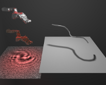
Most of our work on eukaryotes has focused on the development of high-speed holographic microscopy to image the swimming behavior of the malaria parasite Plasmodium berghei. Click the image for more details, including how the holographic microscopy works. If you'd like to have a go at some holographic microscopy yourself, have a look at our downloads page (see navigation bar, above) where you can get our software for free, along with some sample data. The LabVIEW software package and the NI Vision module are both required to run the code.
Magnetic hyperthermia
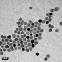
Research from Dr Gonzalo Vallejo Fernandez, lecturer in Physics and Royal Society Industry Fellow, also involves developing innovative methods using magnetic fields to induce hyperthermia in biological tissue.
Data visualisation
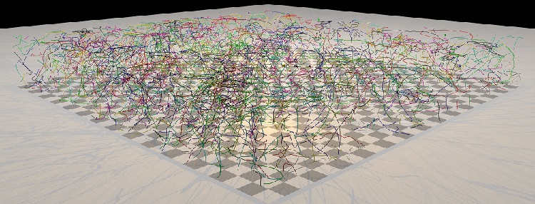
Data visualisation is a big challenge in our work. Because our high-speed cameras generate an enormous amount of information, so are always looking for new ways to display and analyse this information. The page behind this link gives some information about our (rather primitive!) attempts to convey 3D information, and the sort of applications we target.
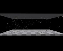
Bacteria and tracking
![]()
We have developed/adapted several techniques for measuring bacterial motility, including a colloid physics technique - Differential Dynamic Microscopy (DDM). This method is analogous to traditional dynamic light scattering measurements, but allows us to measure transport processes (e.g. swimming, Brownian diffusion) on much larger length scales. We applied this in a recent paper to measure collective behaviour in a population, specifically the response to a chemical gradient, or 'chemotaxis'.
Dr Gonzalo Vallejo Fernandez

Dr Gonzalo's research (10) expands over several areas including magnetic thin films for magnetic recording and magnetic nanoparticles for biomedical applications. He has published ~40 peer reviewed papers and is co-inventor in two patents. He has given in excess of 20 oral presentations at international conferences and workshops.
Key Publications:
1. Nat. Commun. 9 5369 (2018) 2. Adv. Funct. Mater. 1706660 (2018) 3. npj Parkinson's Disease 3 34 (2017) 4. Opt. Express 25(23) 28489 (2017) 5 Biophys. J.110(9) 2076 (2016) 6. Malaria J. 15 220 (2016) 7. Nat. Commun. 6 7985 (2015). 8. Proc. Natl. Acad. Sci. USA 111(50) 17771-17776 (2014). 9. Proc. Natl. Acad. Sci. USA 110(47) 18769-18774 (2013). 10. Journal of Physics D (2017).