School of Physics, Engineering and Technology

- School of Physics, Engineering and Technology
- Research and innovation
- Physics of Life
- Next Generation Biosensing
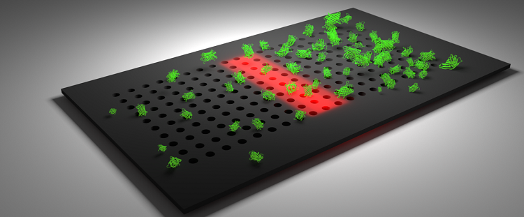
Artist’s impression of a nanophotonic sensor chip
Nanophotonic biosensors
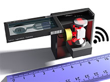
Artist’s impression of the handheld biosensor instrument
- Novel nanophotonic structures and devices can be used for a range of bio-sensing and bio-imaging applications
- The goal is to explore novel photonic concepts, such as metasurfaces and common-path interferometers, for the development of novel sensing and imaging modalities.
- These properties, especially when used in conjunction with suitable microfluidics approaches, can be used to probe interactions of biological molecules, cells, bacteria and tissues with a goal of extracting more information about their function.
- We can now achieve similar or better performance than the laboratory standard enzyme-linked immunoassay (ELISA) method and are working on a handheld instrument that will make this capability accessible to untrained personnel and the public. In particular, we have demonstrated the detection of CRP and procalcitonin at low pg/ml levels [1-3], which are markers that can help discriminate between a bacterial and a viral infection.
The Nanophotonic bio-sensing team is headed by Professor Thomas F Krauss, Chair of Photonics.
Professor Thomas Krauss
The Nanophotonic sensing activity is part of the Photonics research portfolio led by Professor Krauss. The overall aim of the group is to understand and control the light-matter interaction in photonic nanostructures, and to build functional devices that make use of this understanding. Photonic crystals provide an excellent platform for controlling enhanced light-matter interaction, and we are interested in developing them both technologically and conceptually. The thrust of our biologically motivated research is to use the exquisite control over the light field available with photonic crystals to build highly responsive biosensors; in addition, we aim to exploit the strong spatial confinement to build devices that combine high sensitivity with µm-scale spatial resolution, which is unique in the field . Most of the work is based on guided mode resonances [4], which can be understood as nanostructured surfaces that support surface resonances, or simply “resonant surfaces”. Such resonant surfaces allow us to monitor the response of cells and tissue to external stimuli with high spatial and temporal resolution. [5,6]. Much of the activity of the group is now focused on studying bacteria and their susceptibility in the context of antimicrobial resistance and on developing point-of-care healthcare technology devices [7,8].
Funded Projects
“Resonant and shaped photonics for understanding the physical and biomedical world”, Programme Grant, St Andrews, August 2017-July 2022
Light has been used for centuries to image the world around us, and continues to provide profound insights across physics, chemistry, biology, materials science and medicine. However, what are the limits of light as a measurement tool? For example, we can use light to image single bacteria, but can we also use light to trap a single bacterium, identify the bacterial strain and assess its susceptibility to antibiotics? How can we image over multiple length scales, from single cells to multiple cellular tissue, in order to comprehensively map all the neuronal connections in the brain? Can we use a combination of resonance with the wave nature and momentum of light to measure the forces associated with the natural and stimulated motion of a single neuronal cell, or even the extremely small forces associated with phenomena at the classical-quantum interface?
This proposal aims to answer these questions by exploring new and innovative ways in which we can use light to measure the natural world. This research builds on our recent advances in photonics – the science of generating, controlling and detecting light – and in particular will exploit resonant structures and shaped light. These provide us with tools for controlling the interaction of light and matter with exquisite sensitivity and accuracy. We will run three research strands in parallel and by combining their outputs, we aim to address major Global Challenges in antimicrobial resistance, neurodegenerative disease, multimodal functional imaging and next generation force, torque and microrheology.
“Multiparameter Assay for Profiling Susceptibility (MAPS), EPSRC Healthcare Impact Partnership”, September 2017-August 2020
Antimicrobial resistance (AMR) is the ability of microbes to evolve resistance against an antimicrobial treatment. For example, a bacterium can develop resistance to an antibiotic medicine, rendering that medicine ineffective in treating and containing the infection. The loss of effective antibiotics will have a significant impact on our lives, not only increasing the chances of developing a serious infection but also increasing the risk associated with medical procedures. The recent O’Neill review predicts “If we fail to act, we are looking at an almost unthinkable scenario where antibiotics no longer work and we are cast back into the dark ages of medicine”.
While AMR in bacteria occurs naturally over time, the misuse and overuse of antibiotics is accelerating this process. For example, many infections such as tonsillitis are predominantly (80%) viral and can thus not be treated with antibiotics, yet antibiotics are still prescribed. An obvious solution is to introduce new drugs. However, this is not only very costly but it is also inevitable that resistance to any new medicine will develop.
A promising and sustainable solution to the AMR problem is the introduction of diagnostic tests that not only confirm a bacterial infection but also identify the best antibiotic for treating the infection. The aim of this project is to develop a diagnostic that will ensure the right drugs are prescribed at the right time. The technology, called MAPS, is based on silicon photonics. We will exploit this technology to create a diagnostic that will identify the type of bacterium and severity of infection, the presence of resistance mechanisms and the most promising antibiotic for treatment. Working with clinical and industrial collaborators, we will demonstrate and validate the technology for the treatment of urinary tract infections and determine a route for taking it to the market.
“Metalens fluorometer to assess drinking water in Nepal”, EPSRC/GCRF, April 2020-March 2022
The country of Nepal has major issues with the quality of its drinking water. Tools to assess drinking water quality are too expensive and not available. We will address this problem by developing a handheld fluorometer to detect tryptophan-like fluorescence as a proxy for bacterial contamination in drinking water. Our novel design is intrinsically simple and can be made at low cost. The core of the instrument is a high numerical aperture metalens that maximises collection efficiency and that can be made by nanoimprint lithography. The metalens is a nanostructured surface that can impose an arbitrary phase profile to an incoming light beam, and it build on our expertise of making high efficiency metalenses in silicon for operation in the visible wavelength regime [9-11]
The metalens will be designed to be strongly chromatic such that the images for different wavelengths are laterally displaced, which means that the emitter and detector can be placed next to one another. We will develop a translation pathway for prototype instruments to be made in Nepal for use in local communities.
Relevant recent publications
[1] Ahmad Kenaan, Kezheng Li, Isabelle Barth, Steven Johnson, Jie Song and Thomas F. Krauss, “Guided mode resonance sensor for the parallel detection of multiple protein biomarkers in human urine with high sensitivity” accepted, Biosensors & Bioelectronics
[2] D. Conteduca, I. Barth, G. Pitruzzello, C. P. Reardon, E. R. Martins and T. F. Krauss, “Dielectric Fano metasurface for high-resolution near-field sensing and imaging” submitted for publication
[3] Isabelle Barth, Donato Conteduca, Christopher Reardon, Steven Johnson and Thomas F. Krauss, “Common-path interferometric label-free protein sensing with resonant dielectric nanostructures” submitted for publication
[8] Yue Wang, Christopher P. Reardon, Nicholas Read, Stephen Thorpe, Marjan Van Der Woude, Adrian Evans, Neil Todd and Thomas F. Krauss, “Attachment and antibiotic response of early-stage biofilms studied using resonant hyperspectral imaging” submitted for publication
[11] Augusto Martins, Kezheng Li, Juntao Li, Haowen Liang, Donato Conteduca, Ben-Hur V. Borges, Thomas F. Krauss and Emiliano R. Martins, “The “impossible” metalens for wide-field imaging“, submitted for publication