School of Physics, Engineering and Technology
Techniques and Methods
Liquid cell transmission electron microscopy
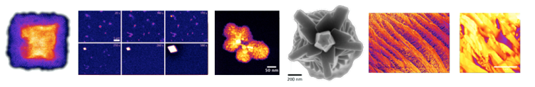
A novel sample holder (Protochips Inc.) for transmission electron microscopy (TEM) allows for the imaging of objects in liquid environments such as minerals forming from supersaturated ionic solutions or biogenic materials that undergo significant changes when dried or frozen to be imaged in TEM. This versatile technique gives access to many aspects of liquid phase processes with an unprecedented spatial resolution of less than 1 nm, especially if scanning TEM (STEM) is used for imaging. We are using this technique to study the dynamics of the formation of minerals such as calcium carbonate or hydroxyapatite from solution. An important aspect in this context is the role of amorphous precursor phases for the growth rates, polymorph selection and shape evolution. Moreover, this method allows for the investigation of biological materials such as bacteria in liquids, which we aim to develop.

Atmospheric scanning electron microscopy
The examination of processes in liquids using a scanning electron microscope (SEM) offers a huge scope of questions to be addressed regarding mineral formation in liquids. Such a unique system (JEOL Clairscope available at the Bioscience Technology Facility), is available in York and consists of an inverted SEM combined with a confocal optical microscope and allows for the imaging of processes and objects in liquids using back-scattered electron detection. The object remains in liquid and under atmospheric condition during the observation. Our studies focus on the precipitation from supersaturated solutions, and in particular the effect of the presence of organic additives - important in many biological systems - on this process. Results of these investigations have been published in the Journal of Structural Biology [9].
Raman microspectroscopy
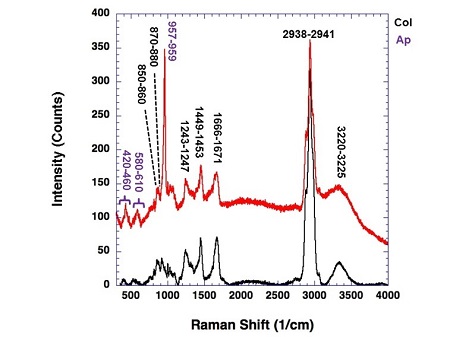
We are using a Horiba XploRA Raman Microscope to study the structure of minerals (e.g. carbonates and phosphate), mineral/organic composites (e.g. corals, bones and teeth) and organic microfibers such as collagen and cellulose. The system is equipped with three laser sources (with wavelengths of 532 nm, 633 nm, 785 nm) allowing for material specific analyses. We have developed in situ stages for Raman spectroscopy allowing to study the stress response of collagen microfibers [5] and the mineralisation dynamics of collagen with hydroxyapatite.
Transmission Electron Microscopy
To view publications on these research areas, please follow this link: Pure pages