School of Physics, Engineering and Technology
Materials
Biomineralisation
The formation of mineral/organic composites such as shells in marine organisms as well as teeth or skeletons is largely determined by the presence of organic additives such as proteins or polysaccharides and the interaction of the ions in solution with these additives prior to precipitation and mineral growth. Novel electron microscopy techniques such as liquid cell transmission electron microscopy or atmospheric scanning electron microscopy allow for the in situ investigation of precipitation processes with unprecedented spatial resolution.
In collaboration with the group of John S. Evans (NYU) we have performed studies of calcium carbonate formation in the presence of the protein AP7 extracted from the nacre of Haliotis Rufensis known to play an active role in the nucleation and crystal growth.
Corals
We show that new insights can be gained on the nano- and microstructure of corallites by TEM investigation of large-scale (15 x 30 µm) FIB lamellae from adult and juvenile scleractinian coral skeletal specimens as shown here:
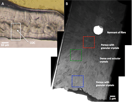
By leaving the FIB prepared lamella within the coral skeletal context (no lift out) the lamella is mechanically more stable and durable while being directly comparable to the larger scale (several tens of microns) not ion-milled skeletal areas by optical analysis. Thus we could identify a crystallographic evolution from a center of calcification outward over acicular and granular (daily) bands. We draw a parallel to the diurnal photosynthetic cycle of the zooxanthellae in symbiotic corals that control the levels of oxygen and carbon availabilities, both recognized as significant drivers of coral calcification processes. The transport of large amounts of glycerol by zooxanthellae and its potential impact on coral calcification was also discussed. This process could play a role in the specific alignment of the aragonite crystals as was previously demonstrated by synthesis experiments with OH group containing additives and the TEM investigation of these precipitates. The juvenile Acropora millepora specimen also showed the large acicular crystals interrupted by thin porous bands, but lacked the nanocrystalline phase, which may be linked to the absence of zooxanthellae and thus the typical daily cycle [9]
Calcifying algae: Rhabdosphaera Clavigera
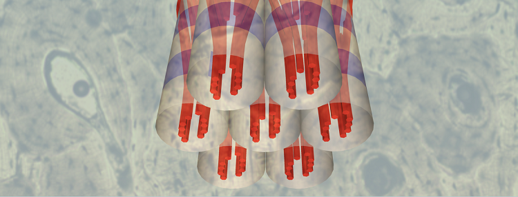
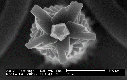
Coccoliths are micrometer scale disks build up from single crystal calcite units, produced by unicellular marine algae, belonging to the phylum Haptophyta. The complex biomineral structure exhibited by these organisms, as depicted here, shows little resemblance to their geological or inorganic equivalents and is impossible to reproduce synthetically:
This implies a stark control by the organism over crystallographic orientation and overall morphology to create functional structures and motivates a detailed study of the microstructure with the aim to unveil fundamental aspects of nanocrystal formation and assembly in biological systems.
Bones and teeth
Cross-section of human bone showing osteons
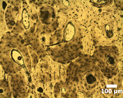
Recently we have commenced to investigate the mineralisation patterns in bones and teeth in collaboration with Paul Genever (Biology, York) and Natalie Reznikov (McGill University, Canada). Aim is to use electron microscopy based techniques to obtain a detailed insight into the correlation between organic phases and the hydroxyapatite phase formed by the bone and tooth forming cells. Here is shown the optical micrograph of human bone sample that had been sliced and thinned down to approx. 10 µm. It shows the osteons and bone cells responsible for the mineralisation. Using electron tomography we succeeded in obtaining a three-dimensional image of the mineral with sub-nanometer resolution and to propose a model of the mineral organisation in the context of the collagen matrix [2].
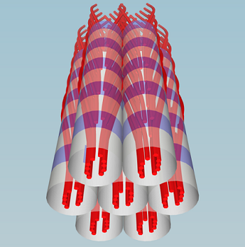
Nanostructure of bone mineral studied with electron tomography in scanning TEM
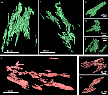
Model of nanocrystal organisation within the collagen matrix of bone
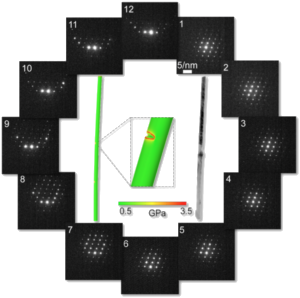
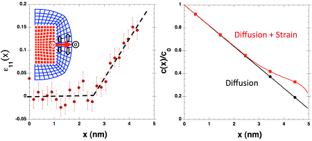
Calcite nanowires
It is generally believed that the formation of minerals in biological systems is strongly determined by the early stages of growth, usually occurring in confinements e.g. created by cells. Therefore, we are particularly interested in the properties of nano-structures such as calcite nanowires grown in confinement. Using electron diffraction in conjunction with finite element calculations (performed by the group of Dr Dorothy Duffy at UCL) we identified an important correlation between the anisotropic crystal structure and lattice distortions such as twist and bending for calcite nanowires of several µm in length and less than 100 nm in diameter:
Nanoparticles for biomedical applications
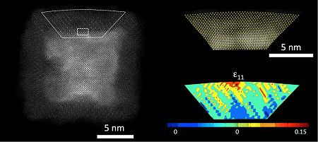
We investigate cluster deposited nanoparticles aimed to be used as nanovectors for theranostic applications. This is a colour-coded high-resolution scanning transmission electron micrograph of a cluster deposited iron particle which has undergone oxidation after exposure to air:
The image was recorded using an aberration-corrected STEM as part of a collaboration with the University of Illinois. A detailed analysis of the lattice strain on the atomic level reveals a significant strain in the oxide layer responsible for an enhancement of the oxidation rate [7].
To view publications on these research areas, please follow this link: Pure Pages