The sensitivity challenge
Scientists in CHyM solve sensitivity problems in scientific and clinical measurements using hyperpolarisation techniques, but why isn’t MRI very sensitive?
MRI and NMR
Magnetic resonance imaging (MRI) and nuclear magnetic resonance (NMR) spectroscopy are extremely powerful techniques used extensively by clinicians and scientists. Despite this, there are fundamental limitations to these methods which scientists in CHyM are seeking solutions to.
MRI is used in hospitals to image the human body, with over 60 million scans performed annually worldwide and in the UK, an average of 41 scans per 1,000 people were performed in 2010. MRI is used to obtain details of the internal structures of the body, especially soft tissue such as muscles, the brain and the heart.
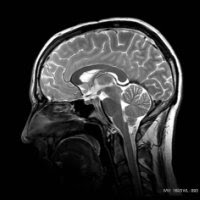
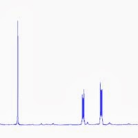
This image shows the internal structure of the human brain, a picture built up of tiny signals from water molecules. NMR spectroscopy is closely related to MRI and is one of the principle tools used by chemists to look at chemical materials. Here, the identity of a compound and details of its properties can be revealed by running simple tests. The second figure shows the NMR spectrum of quinoxaline, where information on the hydrogen atoms in this chemical are represented by sharp peaks.
So what is the problem with MRI and NMR measurements? The answer is: sensitivity.
Low Sensitivity
NMR and MRI measurements are limited because of low sensitivity which leads to poor resolution, contrast and long scan times.
For instance, typical clinical MRI scanners only receive information from 1 in 200,000 molecules that are present in the human body. This would be the equivalent of studying the UK population using only 313 people.
In MRI, this lack of sensitivity restricts the clinician to imaging materials that are present in large proportions in the human body: fat and water. Advanced scanning methods are used to differentiate between these two materials and create levels of contrast that allow internal structures to be revealed.
The following images shows how, despite low signal, contrast between fat and water can be achieved using different scan set-up.
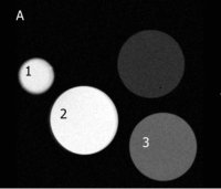
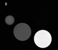
These images show cross sections of 1, a cod-liver oil tablet, 2, a vial of olive oil and 3, a vial of water. Image A uses a particular scan to highlight water (T1 weighted), while image B shows the same tubes but uses a different scan to emphasise fat (T2 weighted). The final vial contains a silicon-based compound as a reference.
Despite achieving this contrast, the low sensitivity of the measurements prevent the clinical observing the complex biochemistry that occurs in the body.
In NMR, low sensitivity means opportunities are missed for observing reaction intermediates, studying reaction mechanisms and studying reaction kinetics.
Low sensitivity has other effects such as long scan times, low resolution imaging, high detection thresholds and expensive equipment.
Solving sensitivity problems
Scientists in CHyM are aiming to solve sensitivity problems in NMR and MRI using hyperpolarisation methods.
General enquiries
- Centre for Hyperpolarisation in MR (CHyM)
Tel: 01904 328886
Further information