Department of Biology

- Department of Biology
- Research and impact
- Molecular and Cellular Medicine
- Milner, Professor Jo
Professor Jo Milner
Emeritus
Overview
Gene expression and disease
Cancer, inflammatory disease, and macular degeneration are all examples of disease states linked with the expression of disease-related genes. One approach for treatment is via selectively silencing the expression of those genes concerned.
RNA interference and siRNA and therapeutics
RNA interference (RNAi) offers an exciting approach for silencing disease-related genes. RNAi is a natural process and is triggered by small interfering dsRNAs (siRNAs). Sequence-specific targeting by siRNA initiates selective destruction of a given mRNA, thereby silencing expression of the gene concerned. siRNAs can readily be designed as therapeutic agents to target given mRNAs, individual mRNA splice variants, or mutant over wild type mRNA.
siRNA is rapidly degraded in the body
One of the major hurdles for the development of RNAi-based therapeutics is the susceptibility of siRNA to degradation by nucleases in the blood stream and in other tissues. In order to counter this problem chemical modification of individual nucleotides within the synthetic siRNA molecule is employed. However, when introduced into mammalian cells, the unnatural nature of the chemically modified siRNA backbone risks activating cellular stress responses. This can result in unwanted side effects of treatment.
Stabilisation of siRNA via single-stranded DNA extension
At York we have explored the possibility of stabilizing siRNA by extension into a single stranded DNA structure known to be resistant to nuclease digestion (Jiang et al., Nucleic Acids Research; 2005). Specifically, one of the siRNA strands (unmodified) was extended with single-stranded DNA (also unmodified). The 3’ terminus of the DNA is composed of the sequence GCGAAGC which forms a compact mini-hairpin structure. In isolation this DNA mini-hairpin is very stable and resistant to nuclease digestion. Remarkably we discovered that the 3’ GCGAAGC (i) does not affect the efficacy of RNA interference, and (ii) confers nuclease resistance to the siRNA. No evidence of cytotoxicity or stress was observed [Allison and Milner, Molecular Therapy Nucleic Acids; 2014].
|
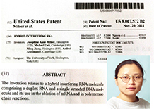
Images of (i) the patent relating to a hybrid interfering RNA molecule comprising siRNA stabilized by a ssDNA extension with 3’GCGAAGC and (ii) Dr. Ming Jiang who was largely responsible for developing this novel technology whilst a member of the YCR P53 Research Unit, Department of Biology, University of York. |
 |
Taking siRNA/GCGAAGC towards the clinic
Synthetic siRNA/GCGAAGC remains (to my knowledge) the only nuclease-resistant siRNA without chemical modification of the RNA backbone. It would appear that the remarkable structural properties of the seven nucleotide sequence GCGAAGC may help unlock the huge potential of RNAi-based therapeutics.
Future development of this novel siRNA/GCGAAGC construct is being taken forward by Argonaute RNA Limited, a UK biotech company in the field of therapeutics.
Profile
Research
| 2010- | Emeritus Professor | Department of Biology, University of York |
| 1991 - 2010 | Professor of Cell Biology, Director YCR P53 Research Unit | Department of Biology, University of York |
| 1972 - 1991 | RA/Senior Research Associate / Merit Award | University of Cambridge |
| 1969 - 1972 | Beit Memorial Research Fellow for Medical Research | University of Cambridge |
| 1969 - 1984 | Fellow, Tutor, College Lecturer in Cell Biology | New Hall College, University of Cambridge |
| 1968 - 1969 | Research Fellow | Harvard Medical School, Harvard University, USA |
| 1968 | PhD | University of Cambridge |
| 1964 | BSc (Zoology) | University of London |
| 1962-1965 | State Scholar |
University of London |
Cambridge University College Fellowship
| 1968 - 1969 | Research Fellow | New Hall |
| 1970 - 1984 | Fellow, Tutor, College Lecturer in Cell Biology | New Hall |
Publications
Cellular p53 : protein structure and functions
1. Milner J. Different forms of p53 detected by monoclonal antibodies in non-dividing and dividing lymphocytes. Nature. 310: 143-145. (1984)
2. *Milner J. and Cook A. The cellular tumour antigen p53: evidence for transformation-related, immunological variants of p53. Virology. 154: 21-30. (1986) 3. Gamble J. and Milner J. Evidence that immunological variants of p53 represent alternative protein conformations. Virology. 162: 452-458. (1988)
4. Cook A. and *Milner J. Evidence for allosteric variants of wild-type p53, a tumour suppressor protein. Br. J. Cancer. 61: 548-552. (1990)
5. *Milner J. and Medcalf E.A. Temperature-dependent switching between "wild-type" and "mutant" forms of p53-Val135. J. Molecular Biology 216: 481-484. (1990)
6. *Milner J. and Watson J.V. Addition of fresh medium induces cell cycle and conformation changes in p53, a tumour suppressor protein. Oncogene 5: 1683-1690. (1990)
7. *Milner J., Medcalf E.A., and Cook A.C. Tumor suppressor p53: analysis of wild-type and mutant p53 complexes. Mol. Cell. Biol. 11: 12-19. (1991)
8. Milner J. The role of p53 in the normal control of cell proliferation. Curr. Opin. Cell Biol. 3: 282-286. (1991)
9. *Milner J. and Medcalf E.A. Cotranslation of activated mutant p53 with wild type drives the wild-type p53 protein into the mutant conformation. Cell 65: 765-774. (1991)
Tumour suppressor p53 is a metal-binding protein and its conformational folding is subject to Hsp70 and redox
10. Hainaut P. and *Milner J. Interaction of heat-shock protein 70 with p53 translated in vitro: evidence for interaction with dimeric p53 and for a role in the regulation of p53 conformation. EMBO J. 11: 3513-3520. (1992)
11. Medcalf E.A., Takahashi T., Chiba I., Minna J., and *Milner J. Temperature-sensitive mutants of p53 associated with human carcinoma of the lung. Oncogene. 7: 71-76. (1992)
12. Hainaut P. and =Milner J. A structural role for metal ions in the "wild-type" conformation of the tumor suppressor protein p53. Cancer Res. 53: 1739-1742. (1993)
13. Hainaut P. and =Milner J. Redox modulation of p53 conformation and sequence-specific DNA binding in vitro. Cancer Res. 53: 4469-4473. (1993)
A conformation hypothesis for p53 protein functions in normal and cancer cells, and implications for anti-cancer therapy
14. Milner J. A conformation hypothesis for the suppressor and promoter functions of p53 in cell growth control and in cancer. Proceedings Royal Society series B 245: 139-145. (1991)
15. Milner J. Forms and functions of p53. Sem. Cancer Biol. 5: 211-219. (1994)
16. Hainaut P., Butcher S., and *Milner J. Temperature sensitivity for conformation is an intrinsic property of wild-type p53. Br. J. Cancer. 71: 227-231. (1995)
17. Milner J. DNA damage, p53 and anticancer therapies. Nature Medicine 1: 879-880. (1995)
18. Milner J. Flexibility: the key to p53 function? Trends Biochem. Sci. 20: 49-51. (1995)
Cellular p53 protein under basal and stress-induced conditions
19. Rubbi C.P. and *Milner J. Non-activated p53 co-localizes with sites of transcription within both the nucleoplasm and the nucleolus. Oncogene. 19: 85-96. (2000)
20. Rubbi C.P. and =Milner J. Analysis of nucleotide excision repair by detection of single-stranded DNA transients. Carcinogenesis. 22: 1789-1796. (2001)
21. Jiang M., Axe T., Holgate R., Rubbi C.P., Okorokov A.L., Mee T., and *Milner J. p53 binds the nuclear matrix in normal cells: binding involves the proline-rich domain of p53 and increases following genotoxic stress. Oncogene. 20: 5449-5458. (2001)
22. Okorokov A.L., Rubbi C.P., Metcalfe S., and *Milner J. The interaction of p53 with the nuclear matrix is mediated by F-actin and modulated by DNA damage. Oncogene. 21: 356-367. (2002)
23. Rubbi C.P. and =Milner J. p53 - guardian of a genome's guardian? Cell Cycle. 2: 20-21. (2003)
24. Rubbi C.P. and =Milner J. p53 is a chromatin accessibility factor for nucleotide excision repair of DNA damage. EMBO J. 22: 975-986. (2003)
25. Allison S.J. and *Milner J. Loss of p53 has site-specific effects on histone H3 modification, including serine 10 phosphorylation important for maintenance of ploidy. Cancer Res. 63: 6674-6679. (2003)
26. Rubbi C.P. and =Milner J. Disruption of the nucleolus mediates stabilization of p53 in response to DNA damage and other stresses. EMBO J. 22: 6068-6077. (2003)
27. Allison S.J. and *Milner J. Remodelling chromatin on a global scale: a novel protective function of p53. Carcinogenesis. 25: 1551-1557. (2004)
28. Warnock L.J., Adamson R., Lynch C.J. and Milner J. Crosstalk between site-specific modifications on p53 and histone H3. Oncogene 27: 1639-1644. (2008)
Cell survival/apoptosis under basal (non-stress) conditions
Apoptosis: the molecular mechanism regulating apoptosis under basal conditions is distinct from stress-induced apoptosis
29. Jiang M. and *Milner J. Bcl-2 constitutively suppresses p53-dependent apoptosis in colorectal cancer cells. Genes and Development 17: 832-837. (2003).
30. Ford J.R., Jiang M., and *Milner J. Cancer-specific functions of SIRT1 enable human epithelial cancer cell growth and survival. Cancer Res. 65: 10457-10463. (2005)
31. Ahmed S U and Milner Jo Basal cancer cell survival involves JNK2 suppression of a novel JNK1/c-Jun/Bcl3 apoptotic network. PLoS One 4 (2009)
32. Milner J. Death without stress. The stiletto approach against disease. The Biochemist. 26: 16-18. (2004)
33. Jiang M. and *Milner J. Selective silencing of viral gene expression in HPV-positive human cervical carcinoma cells treated with siRNA, a primer of RNA interference. Oncogene. 21: 6041-6048. (2002)
33. Milner J. RNA interference for treating cancers caused by viral infection. Expert Opinion Biol. Therapy 3: 459-467. (2003)
Development of siRNA/RNAi technology for novel therapies
34. Jiang M.J. and *Milner J. Gel-based application of siRNA to human epithelial cancer cells induces RNAi-dependent apoptosis. Oligonucleotides. 14: 1-10. (2004)
35. Jiang M., Arzumanov A., Gait M., and *Milner J. A bi-functional siRNA construct induces RNA interference and also primes PCR amplification for its own quantification. Nucleic Acids Res. 33: e151 (2005)
36. Allison S J and Milner Jo. RNA interference by single- and double-stranded siRNA with a DNA extension containing a 3’nuclease-resistant mini-hairpin structure. Molecular Therapy Nucleic Acids (2013) in press.
RECENT PUBLICATIONS
2012
Zahid H. Shah, Shafiq U. Ahmed, Jack R. Ford, Simon J. Allison, John R. P. Knight, and Jo Milner A Deacetylase-Deficient SIRT1 Variant Opposes Full-Length SIRT1 in Regulating Tumor Suppressor p53 and Governs Expression of Cancer-Related Genes. Molecular & Cellular Biology 32; 704-716 (2012)
Warnock L J, Raines, S A and Milner Jo Aurora A mediates cross-talk between N- and C-terminal post-translational modifications of p53. Cancer Biology and Therapy 12; 1059-1068 (2012)
Knight J R P K and Milner Jo SIRT1 metabolism and cancer. Current Opinion in Oncology ; 24, 68-75 (2012).
2013
Knight, John R P., Willis, Anne E. and Milner Jo Active regulator of SIRT1 is required for ribosome biogenesis and function. Nucleic Acids Research ; 41(7), 4185-4197 (2013)
Knight JRP, Allison SJ and Milner Jo Active regulator of SIRT1 is required for cancer cell survival but not for SIRT1 activity. Open Biology ; 3, 130130 (2013)
Allison S J and Milner Jo RNA interference by single- and double-stranded siRNA with a DNA extension containing a 3’nuclease-resistant mini-hairpin structure. Molecular Therapy Nucleic Acids (2013) in press.
Current Activities
2010 – 2015: In addition to completing publications arising out her YCR-funded research in the Department of Biology, Jo Milner has presented this work at USA Keystone Conferences on Aging and Diseases of Aging (2012) and The Biology of the Sirtuins (2015).
External PhD examiner (i) Birkbeck/UCL, London University in 2015, and (ii) the Faculty of Life Sciences at The University of Manchester in 2016.
| 2017: | |
| Founder and Sole Director: | ChimeraDNA Ltd, developing synthetic biology and nanotherapeutics |
| Award: | Biorenewables Business Assistance Project Award, European Regional Development Fund (ERDF). £1,224 |
| Catapult & Rainbow Seed Fund |
Invitee: |
Current activities:
Acting Chief Scientific Officer for a newly founded UK biotech company in the field of biomedicine. As sole scientist this informal role has included:-
- development of the company’s scientific rationale and projected translation towards the clinic,
- strategy for this purpose
- identification of and meetings with specialist advisors to the company
- Company board meetings in London and in Cambridge
Design, management and interpretation of a research project carried out by Horizon Discovery, Cambridge. The project was successful and, using novel technologies, confirmed previous results arising out of Jo Milner’s YCR-funded research in the Department of Biology