Our people
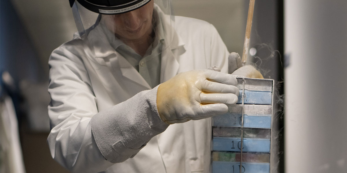
The Jack Birch Unit brings together researchers from disciplines and departments across the University of York.
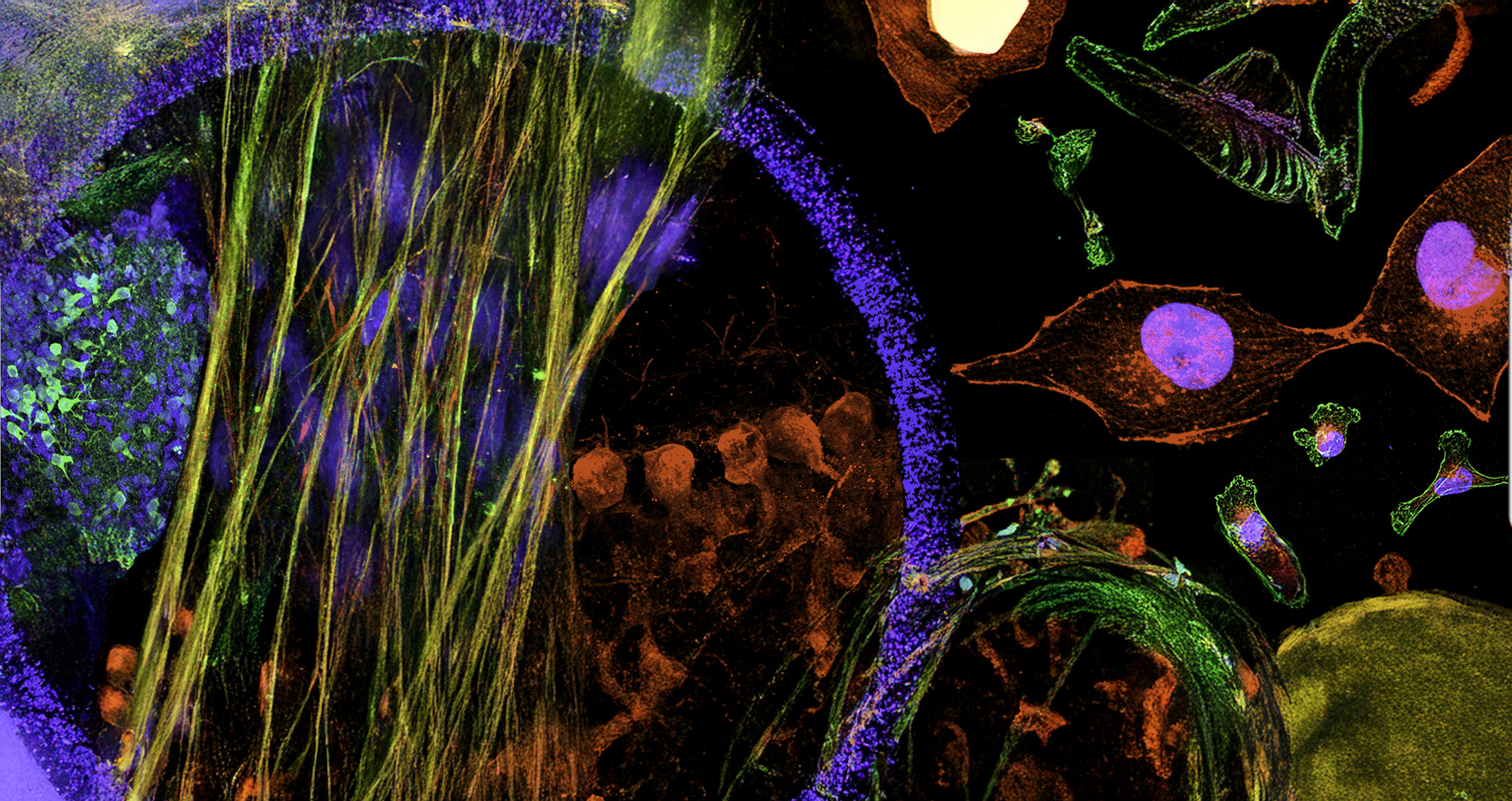
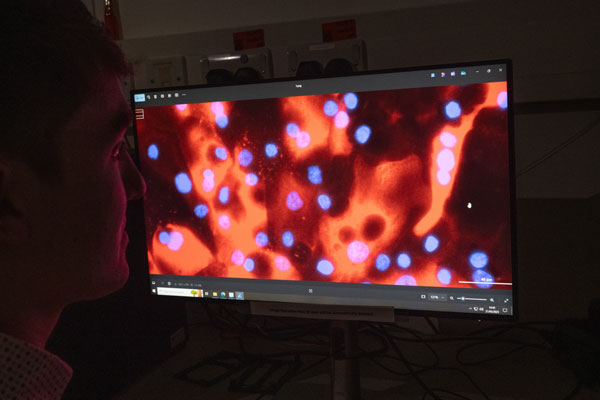
Pioneering cutting-edge cancer research at the University of York.

“Our funding from York Against Cancer presents a fantastic opportunity to expand our research in breast, bladder, ovary, colon and kidney cancers to accelerate the development of new treatments that can make a real difference to the lives of cancer patients in the future.”
Professor Will Brackenbury, Director of the Jack Birch Unit.
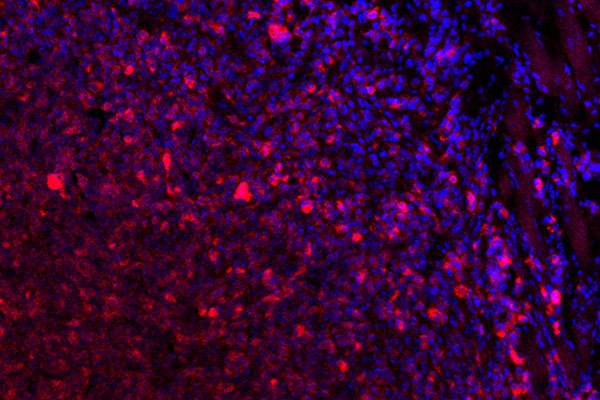
The Jack Birch Unit is leading research into poorly understood aspects of the local microenvironment of epithelial tumours.
As part of the York Biomedical Research Institute and in collaboration with the Hull York Medical School, our strategy is built around building on our strong track record in fundamental bladder and breast cancer research, whilst growing capabilities in other tumour types, including those of the ovary, colon and kidney, as well as undertaking translational research to move findings towards the clinic.