
Dr Adrian Mountford, Department of Biology, University of York, PO Box 373, York YO10 5YW, UK
Telephone: +44(0) 1904 328595 - Email: apm10@york.ac.uk
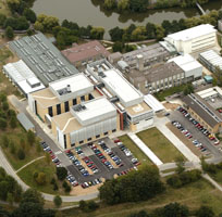
The Department of Biology,
University of York, UK.
The Department of Biology at the University of York is one of the UK's leading biological sciences departments. With approximately 60 academic staff, it covers the spectrum of contemporary biological sciences from molecular aspects of cancer to field ecology. All aspects of activity are highly rated in peer review: teaching was scored at 24/24 by the Quality Assurance Agency (QAA) for higher education, and research rated at "5" in the most recent national Research Assessment Exercise (RAE).
The Biology Department was founded in 1965 as an integrated and multidisciplinary unit. Traditional boundaries that fragment the subject of biology are absent, and as a result a number of highly productive interdisciplinary collaborations have emerged. In 2002, new laboratories were constructed through a £21.6M Joint Infrastructure Funding award from BBSRC, with additional funds from Yorkshire Cancer Research, HEFCE and the University of York. The labs provide 10,000m2 of research space, as well as the Structural Biology Laboratory of the Department of Chemistry.
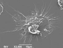
Scanning electron micrograph of activated
dendritic cell with characteristic
‘spiked’ morphology
The Department of Biology also has a Technology Facility that enables researchers to gain access to state-of-the-art equipment and the associated expertise to use such equipment effectively. The Technology Facility is centrally located within the Department and has some 2,000m2 of laboratory space, £6.6 million of new equipment, and 19 expert staff members. It is organised into six specialist laboratories, each led by an experienced technologist with trained technical support.
The Schistosome Research Group
The group focuses on how certain parasitic helminths (i.e. Schistosoma and Trichobilharzia) trigger the early innate immune response, particularly at the skin site of exposure. We have recently developed protocols to label infectious cercariae with fluorescent dyes in order to visualise the larvae as they penetrate the host skin, and to be able to identify which types of cells take up the labelled antigens by phagocytosis or endocytosis.
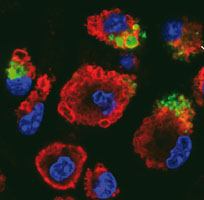
Activated macrophages labelled with
Lamp-1 (red), DAPI (blue) taking up CFSE
labelledparasite material.
The innate inflammatory response in the skin is co-ordinated by a cascade of cytokines and chemokines, with IL-12 and IL-10 important mediators of cross-regulation and the induction of Th1 versus Th2-type responsiveness. However, exposure to multiple doses of infectious cercariae leads to a state of immune hyporesponsiveness that may be related to the development of antigen presenting cells that have an ‘alternative activation’ status. Analysis of dendritic cells has shown that molecules released by the invading parasite are stimulants of their maturation, and that primed dendritic cells induced polarised Th2-type responses in vivo. Proteomic technologies are currently in use to define surface or secreted markers of Th2-inducing dendritic cells.
Current group members:
| Dr Adrian Mountford (Group leader) Dr Joe Turner (Postdoctoral Fellow) Dr Fabienne Callion (Postdoctoral Fellow) |
Ross Paveley (Postgraduate student) Sarah Aynsley (Postgraduate student) Peter Cook (Postgraduate student) |