Dr Aneurin J Kennerley
Lecturer in Chemistry (Magnetic Resonance Imaging)
+44 (0)1904 324230
Email: aneurin.kennerley@york.ac.uk
Twitter: @MagneticDR_K
Research
Basically as a ‘methods development’ group we have lots and lots of fun pushing imaging techniques like MRI to their very limits. We work primarily at 3 Tesla (Human), 7T (pre-clinical) and 9.4T (samples). We use SIEMENS and BRUKER based MR systems. If you want help with anything MRI based – get in touch via email aneurin.kennerley@york.ac.uk

My biomedical imaging research works towards answering important and clinically relevant questions, be that in neuroimaging (particularly functional MRI), Oncology, Cardiovascular research, Musculoskeletal or Liver/Gastrointestinal medicine. Unfortunately, even though use is wide ranging, in the majority of cases an in-depth understanding of the complex MRI signal source (in terms of the underlying physics, chemistry and biology) is lacking. I address this by utilising novel multi-model imaging approaches; using data from a combination of imaging techniques to parameterise biophysical models. In turn, data from these models can feedback to the imaging laboratory helping further develop and refine these techniques. As a member of the Centre for Hyperpolarised Magnetic Resonance I also explore novel physical chemistry to push the signal detection limits. The above approaches help constantly push (push constantly; is that a split infinitive Mr Spock?) MR for improved diagnostics and novel therapeutics.
Examples of ongoing projects are highlighted below.
Current Funding
- Feb 2021 - Jun 2021: £36,000; EPSRC IAA. “A RAY OF LIGHT AGAINST AGE RELATED NEURODEGENERATION: Light transport modelling & NMR metabolomics for quantification of the protective effects of direct photobiomodulation” – Co-Investigator (collaboration with Dr Heidi Baseler, HYMS)
- Jul 2020 – Dec 2021: £70,000; EPSRC IAA. “Improving in-vivo imaging capability of SABRE through X-nuclei detection” – Principle Investigator (collaboration with Professor Simon Duckett, Chemistry & Professor Sven Plein, University of Leeds)
- Jul 2020 – Dec 2021: £25,000; EPSRC IAA. “Super-Powering Diagnostic Sodium MRI to Understand Breast Cancer Heterogeneity” – Co-Investigator (collaboration with Dr William Brackenbury, Biology & Dr Simon Bale, Engineering)
- Oct 2019 – Dec 2021: €40,000; York-Maastricht Partnership. “Development of an integrated MR imaging acquisition and analysis platform” – Principle Investigator (collaboration with Dr Laurentius Huber, MU)
- Jun 2018 – Dec 2021: £299,000; CRUK: “Harnessing the ionic microenvironment as a novel diagnostic and treatment approach for breast cancer” - Co-Investigator (collaboration with Dr William Brackenbury, Biology)
- Jul 2017 – July 2021: £492,000; CRUK: “Magnetic Resonance Targeting of novel oncolytic viruses to tumours: Investigating Efficacy & Modelling forces” - Co-Investigator (collaboration with Dr Munitta Muthana, University of Sheffield)
Neuroimaging Research
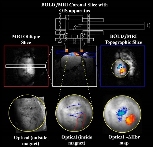
My preclinical research focuses on developing multi-modal imaging technology (MRI and intrinsic optical imaging spectroscopy) to investigate the underlying haemodynamic mechanisms of the functional (f)MRI Blood Oxygenation Level Dependent (BOLD) signal.
A)
B)
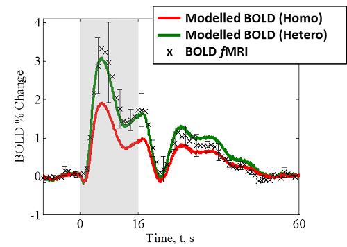
Figure 1 – Development of multi-modal fMRI and optical imaging; A) methods involved; B) using MCS to predict BOLD fMRI signal from the haemodynamics with a basic homogeneous tissue model and MRI parameterised heterogeneous tissue model. The latter shows a better fit to experimental data.
I am currently working on improvements to the optical recording from brain – implanting spatial frequency domain imaging in preclinical models to estimate changes in scattering which can be attributed to cellular swelling during activity. This imaging technique will be combined with diffusion weighted fMRI which is also related to microstructural changes in cell structure. We collect supporting diffusion weighted fMRI data in human at the York Neuro-imaging Centre.
An arm of this research also concerns the laminar based information that can be captured with high resolution fMRI.
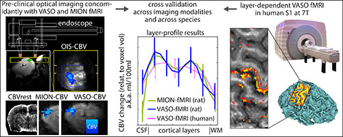
Figure 2 – We utilise concurrent fMRI and optical measures to explore the exact contrast mechanisms of layer-dependent VASO fMRI. Quantified cortical layer profiling is demonstrated and in agreement between both VASO and contrast enhanced fMRI (using monocrystalline iron oxide nanoparticles, MION) in rodent models. Responses show high spatial localisation to layers of cortical excitatory and inhibitory processing independent of confounding large draining veins which hamper BOLD fMRI studies. While we find increased VASO based CBV reactivity (3.1 ± 1.2 fold increase) in humans compared to rats it is demonstrated that this reflects differences in stimulus design rather than confounds of the VASO signal source.
The BOLD fMRI signal is not a direct measure of neuronal activity. It is a confound of the haemodynamic response, in turn driven by neuronal activity. Using X-nuclei NMR we can measure metabolic activity directly, targeting important molecules like adenosine triphosphate (ATP). Our current research is pushing magnetisation transfer based 31P MRS to help us understand mitochondrial function in health, disease and in response to novel treatment.
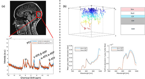
Figure 3 – Preliminary (a) non-localised 31P magnetisation transfer MRS measurements from the healthy human brain. Magnetisation transfer through PCr/ATP exchange causes a quantifiable decrease in the PCr peak integral as a marker of cellular metabolism in-vivo. (b) All MRS data will be correlated to estimates of cytochrome-c light absorption determined from parameterised Monte Carlo simulations of light transport.
Hyperpolarisation Research
To improve diagnostic capability of MRI, increased sensitivity to atypical activity of biomolecules associated with disease progression is urgently required. Their inherent low abundance makes measurement with standard MRI problematic. ‘Hyperpolarised’ contrast agents permitting detection of such biomolecules presents as a novel solution. At the University-of-York we empower a faster/cheaper hyperpolarisation method known as signal amplification by reversible exchange (SABRE). We are now moving towards X-nuclei detection and improvements in solvent approach to propel this ‘next-generation’ technology towards clinical uptake; building foundations needed to deliver real patient impact. Target metabolites include pyruvate to link into existing Dynamic Nuclear polarisation (DNP) research and our ongoing research efforts in the field of oncology.
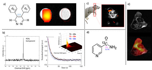
Figure 4 – Recent developments towards in-vivo 13C based SABRE hyperpolarisation detection. (a) Target substrate 4,5-di(phenyl-d5)-3,6-d2-pyridazine has been optimised to 25% polarisation in DCM. Subsequent phantom based Chemical Shift Image (CSI) detection utilised a preclinical 7T MRI system with dual tuned 1H/13C 72mm ID volume coil. 13C image superimposed onto 1H structural image. 13C CSI image integrated over detectable peak @ 136ppm in resultant spectrum (b). FID based signal decay with 10o flip angle used to characterise T1 lifetime of 22s in agreement with published literature.
Oncology Research
We also push X-nuclei MRI development in the field of oncology. Our research shows that Na+ plays a vital role in disease progression, making non-invasive 23Na-MRI an informative diagnostic tool. Indeed, our research in this field is proving that 23Na MR detection/imaging can be an improved indicator of breast cancer as routine 1H diffusion MR measures. We continue to develop this technique using improved detection antennae (with collaborators in Engineering).

Figure 5 - 23Na MRI for assessing mammary tumour ionic microenvironment in mice. (a) 23Na MR scans confirm raised Na+ in tumour tissue with longitudinal assessment showing significantly lower Na+ after treatment with chemotherapy. (b) Hypoxia mimicked with the HIF stabiliser DMOG (1 µM) in breast cancer cells shows increased intracellular Na+ (using indicator SBFI-AM) over controls. Sodium-potassium ion pump inhibitor (Ouabain, 50mM) used as a positive control. (c) 3.5 fold theoretical gain in SNR possible through cooling single RF coil loop with liquid N2 (77K). Increase in SNR can be used to increase resolution & probe response heterogeneity. (d) Apparent diffusion coefficient (ADC) MRI (standard clinical approach for diagnosing cancer) compared with 23Na MRI. AUR-ROC plot (and confusion matrices - not shown) confirm similar detection accuracy between thermal 23Na-MRI and ADC. Detection accuracy improves if both metrics are considered.
Novel drug delivery mechanisms are currently a hot topic; a recent successful multidisciplinary research project I was profoundly involved in used Iron tagged macrophages to carry anti-cancer drugs to tumour sites within the body. I have shown by in vitro and in vivo that by pulsing the magnetic field gradients on an MRI system we can steer these labelled macrophages in a given direction. Thus when gradient targeting is applied towards tumour sites there is an increased uptake of the macrophage cells (upwards of 800% increase) and so the drug for improved therapy.
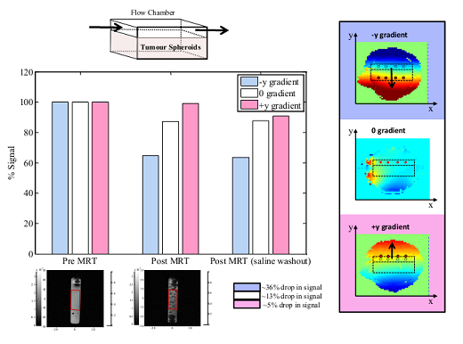
Figure 6 – in-vitro demonstration of magnetic resonance targeting. The MR imaging gradients can be pulsed in a given direction to effectively ‘steer’ iron labelled particles in that direction. This can lead to a significant uptake of particles into a specific area – for example a tumour site to deliver therapy.
Real Time MRI
The fast switching gradients on modern MRI scanners, alongside iterative image reconstruction, now permit deployment of real time imaging methodologies. Alongside important cardiac applications, real time (RT) MRI offers new opportunities for the non-invasive functional monitoring of the mechanics of speech, swallowing and breathing. Quantitative monitoring of such mechanics finds relevance before and after major ablative/reconstructive maxillofacial surgery. Many patients, in particular those with head/neck cancer or major trauma, have serious enduring post-operative problems; e.g. difficulty in swallowing and speech. These long-term problems lead to malnutrition, isolation and depression. RT MRI methods in this context can improve surgical planning, post-operative short and long term rehabilitation and monitoring. Furthermore, RT-MRI can provide insight into adaption following major surgery in this region. In collaboration with Angelika Sebald (Computer science) we are assessing the safety/feasibility of RT-MRI and establishing protocols for the evaluation of swallowing and speech. Data from this unique monitoring methodology will help improve the current less than optimum post-treatment support and patient management; ultimately improving the quality of life of maxillofacial patients.
Some of our real time imaging work is showcased on the following public information site aimed at maxillofacial patients and carers: Maxfacts.uk
News
- 12th June 2021: This is the planned date for our return to bringing our science to you! We will be part of the York Festival of Ideas. Join us for an interactive and entertaining show mixing magic and world leading science, and see if we can read your mind LIVE with our cardboard brain scanner! Intrigued? You should be. For more information, check out the Festival of Ideas website.
- 12th April 2021: We expect to start scanning volunteers again on our 3T MRI system. If you are interested in helping us develop MR based methods please get in touch.
- February 2021: More EPSRC IAA funding. This time for continuing our 31P MRS project looking at ATP flux in the brain pre- and post-red light treatment. Watch this space. Elizabeth Fear will be leading on this project!
- January 2021: A big welcome to Gulmira – who joins us from Kazakhstan to complete an MSc in SABRE hyperpolarisation. She will work on characterising hyperpolarisation of Pyruvate.
- November 2020: We are back in the 7T MRI facility at York pushing the frontiers of fMRI research. This project is led by our keen PhD student Frida Torkelsen. Let's hope for less disruption moving forwards.
- 12th November 2020: Another week another Dr from CHyM @ChemistryatYork successfully defending her thesis on SABRE Hyperpolarisation. Congratulations from all of us Dr Jenny Lewis.
- 30th October 2020: Congratulations to Dr Elizabeth J Fear (@ElizabethFear2) - who successfully defended her PhD thesis ‘An integrated approach to Hyperpolarised Magnetic Resonance’ yesterday @chemistryatYork. A huge well done from all of us at CHyM. Well deserved. External: Professor Archibald (Hull). Look how happy she is!
- August 2020: We have secured ESPRC IAA funding to push SABRE hyperpolarisation into X-nuclei detection. This will help us push the innovative technology forward towards in-vivo detection, where there are no confounds from endogenous signal contrast. Watch this space
- August 2020: We present 3 abstracts at the virtual ISMRM meeting
- March 2020: We are closing the lab to help project our researchers from the COVID-19 pandemic. Stay safe everyone.
- February 2020: Elizabeth Fear wins a travel award to present our novel hyperpolarisation research at the 2020 ISMRM in Sydney Australia.
Publications
H-index = 22; i-10 index = 34 (as of 23rd March 2021)
Visit Aneurin's Google Scholar page for a full list of publications.
2022
- James, AD., Leslie, T.K., Kaggie, JD., Wiggins, L., Patten, L., O’Duinn, J.M., Langer, S., Labarthe-Last, M.C., Riemer, F., Baxter, G., McLean, MA., Gilbert, FJ., Kennerley, AJ., Brackenbury WJ. (2021) Sodium accumulation in breast cancer predicts malignancy and treatment response. British Journal of Cancer, 1-13 doi.org/10.1101/2021.04.14.439494
- Kennerley, AJ., Mitchell, DA., Sebald, A., Watson, I. (2021) Real-time magnetic resonance imaging: mechanics of oral and facial function. British Journal of Oral and Maxillofacial Surgery. doi.org/10.1016/j.bjoms.2021.10.008
- Fear, EJ., Kennerley, AJ., Rayner, PJ., Norcott, P., Roy, SS., Duckett, SB., (2022) SABRE Hyperpolarised Anti-Cancer Agents for use in 1H MRI. Mag Res Med. 88(1), 11-27. doi.org/10.1002/mrm.29166
- Howard, F., El-Janabi, H., Staniland, S., Kennerley, A.J., Patel, P., Collado-Rojas, C., Allwood, D., Smith, E., Vadakekolathu, J., Pockley, G., Connor, J., Muthana, M. (2022) Nanobugs as Drugs – Bacterial Derived Nanomagnets Enhance Tumour Targeting and Oncolytic Activity of HSV-1 Virus. Small. 2104763 doi.org/10.1002/smll.202104763
- Rayner, P.J., Fekete, M., Gater, C.A, Ahwal, F., Turner, N., Kennerley, A.J. & Duckett, S.B. (2022) Real-Time High-Sensitivity Reacation Monitoring of Important Nitrogen-Cycle Synthons by 15N Hyperpolarised Nuclear Magnetic Resonance. J. Am. Chem. Soc. 144, 8756-69
2021
- Huber, L., Poser, BA., Kaas, AL., Fear, EJ., Besbach, S., Berwick, J., Goebel, R., Turner, R. & Kennerley, AJ. (2021) Validating layer-specific VASO across species. NeuroImage, 118195
- Plesia, M., Stevens, O., Gavin, L., Kendall, C., Coldicott, I., Kennerley, A.J., Miller, G., Shaw, P., Mead, R., Day, J., Alix, J. (2021) In vivo Fibre Optic Raman spectroscopy of Muscle in Preclinical Models of Amyotrophic Lateral Sclerosis and Duchenne Muscular Dystrophy. ACS Chemical Neuroscience 12(10), 1768-76
2019
- Lali, W., Roy, SS., Tickner, BJ., Ahwal, F., Kennerley, AJ., Duckett, SB. (2019) Hyperpolarising pyruvate through signal amplification by reversible exchange (SABRE); Angewandte Chemie 131 (30), 10377-10381
- Leslie, TK., James, AD., Zaccagna, F., Grist, JT., Deen, S., Kennerley, AJ.,Riemer, F., Kaggie, JD., Gallagher, FA., Gilbert, FJ., Brackenbury, WJ. (2019) Sodium homeostasis in the tumour microenvironment. Biochimica et Biophysica Acta (BBA)-Reviews on Cancer 1872 (2), 188304.
2017
- Bruyns-Haylett, M., Luo, J., Kennerley, A.J., Harris, S., Boorman,L., Milne, E., Vautrelle, N., Hayashi, Y., Whalley, B.J., Jones, M., Berwick, J., Riera, J., Zheng, Y. (2017) The neurogenesis of P1 and N1: A concurrent EEG/LFP study, NeuroImage 146, 575-88.
- Dounavi, M.E., Selvarajah, D., Tesfaye, S., Martin, C., Kennerley, A.J., Wilkinson, I.D. (2017) Evaluation of cerebral perfusion in Type 2 diabetes using magnetic resonance imaging. Diabetic Medicine, 34. 48-9.
- Harris, S., Boorman, L., Kennerley, A.J., Sharp, P., Martin, S., Redgrave, P. Schwartz, T., Berwick, J. (2017) Seizure epicenter depth and translaminar field potential synchrony underlie complex variations in tissue oxygenation during ictal initiation. bioRxiv, 172148.
- Harris, S., Boorman, L., Das, D., Kennerley, A.J., Sharp, P., Martin, S., Redgrave, P. Schwartz, T., Berwick, J. (2017) Physiological and pathological brain activation in the anesthetized rat produces hemodynamic-dependent cortical temperature increases that can confound the BOLD fMRI signal. bioRxiv, 205609.
- Hewitt, B., Yap, MH., Hodson-Tole, EF., Kennerley, AJ., Sharp, PS., Grant, RA. (2017) A novel automated rodent tracker (ART), demonstrated in a mouse model of amyotrophic lateral sclerosis, Journal of Neuroscience methods 300, 147-156.
2016
- Dall’Ara, E., Boudiffa, M., Taylor, C., Schug, D., Fiegle, E., Kennerley, A.J., Damianou, C., Tozer, G.M., Kiessling, F., Muller, R. (2016) Longitudinal imaging of the aging mouse. Mechanisms of Ageing and Development, 160, 93-116.
- Kazan, SM., Mohammadi, S., Callaghan, MF., Flandin, G., Huber, L., Leech, R., Kennerley, A., Windischberger, C., Weiskopf, N. (2016) Vascular Autocalibration of fMRI (VasA fMRI) Improved Sensitvity of Population Studies: A Pilot Study. NeuroImage 124, 794-805
- Slack, R., Boorman, L., Patel, P., Harris, S., Bruyns-Haylett, M., Kennerley, A.J., Jones, M., Berwick, J. (2016) A novel method for classifying cortical state to identify the accompanying changes in cerebral hemodynamics, Journal of Neuroscience methods, 267, 21-34
2015
- J Bain, L Ruiz-Perez, AJ Kennerley, S Muench, R Thompson, G Battaglia, SS Staniland (2015) In situ formation of magnetopolymersomes via electroporation for MRI. Nano Letters. Scientific Reports 5.
- L Boorman, S Harris, M Bruyns-Haylett, A Kennerley, M Jones, Y Zheng, P Redgrave, J Berwick (2015) Long-latency reductions in gamma power predict negative hemodynamics that underlie the negative BOLD signal. Journal of Neuroscience. 35(11) 4641-56
- L Huber, J Goense, A Kennerley, R Trampel, M Guidi, E Reimer, D Ivanov, N Neef, C.J. Gauthier, R Turner, and H. E. Möller (2015) Cortical lamina-dependent blood volume changes in human brain at 7T. NeuroImage 107, 23-33
- A J Kennerley*, M Muthana*, R Hughes, E Fagnano, J Richardson, M Paul, C Murdoch, F Wright, C Payne, M Lythgoe, N Farrow, J Dobson, J Conner, J M Wild, C Lewis (2015) Directing Cell Therapy to Anatomic Target Sites in-vivo with Magnetic Resonance Targeting. Nature Coms 6.
- M Muthana, A J Kennerley, E Murphy, R Hughes, J Conner, F Wright, M Lythgoe, J Dobson, J M Wild, C Lewis (2015) Use of Magnetic Resonance Targeting to Steer OV-Loaded Cell Based Therapies to Tumour Sites in-vivo. Journal of immunotherapy of cancer 3(2) 1.
- Sharp, P., Kennerley, A.J., Boorman, L., Harris, S., Azzouz, M., Berwick, J. (2015) The Importance of macroscale optical imaging spectroscopy in a mouse model of neurovascular coupling. Scientific Reports 5.
2014
- RA Grant, P Sharp, AJ Kennerley, J Berwick, A Grierson, T Ramesh, TJ Prescott (2014) Abnormalities in whisking behaviour are associated with lesions in brain stem nuclei in a mouse model of amyotrophic lateral sclerosis. Behavioural Brain Research 259. 274-83
- S Harris, L Boorman, Y Zheng, A Kennerley, M Bruyns-Haylett, P.G. Overton, H. Ma, M. Zhao2, T.H. Schwartz, J Berwick (2014) Coupling between gamma-band power and cerebral blood volume during recurrent acute neocortical seizures. NeuroImage (97) 62-70
- S Harris, L Boorman, M Bruyns-Haylett, A Kennerley, H. Ma, M. Zhao, P.G. Overton, T.H. Schwartz, J Berwick (2014) Contralateral dissociation between neural activity and cerebral blood volume during recurrent acute focal neocortical seizures. Epilepsia 55(9) 1423-30
- L Huber, J Goense, A Kennerley, D Ivanov, S N. Krieger, J Lepsien, R Trampel, R Turner, and H. E. Möller (2014) Investigation of the neurovascular coupling in positive and negative BOLD responses in human brain at 7T. NeuroImage 97: 349-62
- AJ Kennerley, L Boorman, S Harris, J Berwick (2014) Simultaneous Functional Magnetic Resonance and Two-Dimensional Optical Imaging Spectroscopy. Neurovascular Coupling Methods, Springer New York 3-20
- E.E. Vidal-Rosas, S.A. Billings, Y Zheng, J.E.W. Mayhew, D Johnston, A Kennerley, D. Coca (2014) Reduced order modelling of light transport in tissue for real-time monitoring of brain haemodynamics using diffuse optical tomography. Journal of Biomedical Optics. 19(2) 026008
2013
- Harris, S., Boorman, L., Kennerley, A., Bruyns-Haylett, M., Overton, P.G., Ma, H., Zhao, M., Schwartz, T.H., Berwick, J. (2013) Linear coupling between synchronized gamma oscillations and cerebral blood volume during recurrent focal neocortical seizures. Journal of Cerebral Blood Flow & Metabolism. 33:1595-604.
- Kennerley, A.J. (2013) Understanding Neurovascular Coupling: Are you BOLD enough? Proceedings in British Journal of Neurosurgery, 27(3) pg 287
- Martin, C.J., Kennerley, A.J., Berwick, J., Mayhew, J.E.W. (2013) Functional Magnetic Resonance Imaging in conscious rats using a chronically implanted surface coil. J Mag Res Im 38(3) 739-44.
2012
- Devonshire, I.M., Papadakis, N.G., Port, M., Berwick, J., Kennerley, A.J., Mayhew, J.E.W., Overton, P.G. (2012) Neuro-vascular coupling is brain region-dependent. Neuroimage 59(3):1997-2006
- Kazan, S., Reynolds, S., Bluff, J., Kennerley, A.J., Wholey, E., Berwick, J., Cunningham, V., Paley, M. Tozer, G., (2012) Kinetic modeling of hyperpolarized 13C Pyruvate pyruvate and Lactate lactate metabolism in tumours using a measured arterial input function. Mag Res Med. DOI: 10.1002/mrm.24546.
- Kennerley, A.J., Harris, S., Bruyns-Haylett, M., Boorman,L., Zheng, Y., Berwick, J. (2012) Early and late hemodynamic responses provide insight into the neurogenic nature of neurovascular coupling. Journal of Cerebral Blood Flow & Metabolism, 32, 468–480
- Kennerley, A.J., Boorman, L., Johnston,D., Port, M., Zheng, Y., Mayhew, J.E., Berwick, J. (2012) Concurrent high field (7T) fMRI with 2D Optical Imaging Spectroscopy: Investigation of the haemodynamics underlying the BOLD signal. NeuroImage, 61 (1) , pp. 10-20
- Zheng, Y., Luo, J., Harris, S., Kennerley, A., Berwick, J., Billings, S., Mayhew, J.E. (2012) Balanced excitation and inhibition: model based analysis of local field potentials. NeuroImage. 63(1). Pg. 81-94
2011
- Lawrie, A., Hameed, A.G., Chamberlain, J., Kennerley, A.J., Arnold, N., Crossman, D., Francis, S. (2011) Paigen diet-fed apolipoprotein E knockout mice develop severe pulmonary hypertension in an interleukin-1-dependent manner. Am J Path, 179(4):1693-705.
- Mead, R.J., Bennett, E.J., Kennerley, A.J., Sharp, P., Sunyach, C., Kasher, P., Berwick, J., Pettmann, B., Battaglia, G., Azzouz, M., Grierson, A., Shaw, P.J. (2011) Optimised and Rapid Pre-clinical Screening in the SOD1G93A Transgenic Mouse Model of Amyotrophic Lateral Sclerosis (ALS). PLoS ONE 6(8): e23244
2010
- Zheng, Y., Pan, Y., Harris, S., Billings, S., Coca, D., Berwick, J., Kennerley, A.J., Johnston, D., Mayhew, J.E. (2010) A mathematical model of neurovascular coupling suggesting both vasodilation and vasoconstriction. J Cereb. Blood. Flow. Metab. 29: S19-20.
- Boorman, L.W., Kennerley, A.J., Johnston, D., Jones, M., Martin, C.J., Zheng, Y., Mayhew, J.E., Berwick, J. (2010) Negative Blood Oxygen Level Dependence in the rat: A model for investigating the role of suppression in neurovascular coupling. Journal of Neuroscience. 30(12): 4285-94
- Kennerley, A.J., Mayhew, J.E., Berwick, J. (2010) Vascular Weightings of BOLD and CBV fMRI signals: A direct comparison of statistical mapping and histological sections. The Open NeuroImaging Journal. 4:1-8
- Zheng, Y., Pan, Y., Harris, S., Billings, S., Coca, D., Berwick, J., Jones, M., Kennerley, A.J., Johnston, D., Martin, C., Devonshire, I.M., Mayhew, J.E. (2010) A dynamic model of neurovascular coupling: implications for blood vessel dilation and constriction. NeuroImage 52:p1135–1147
2009
- Bonesi, M., Matcher, S., Kennerley, A.J., Meglinski, I. (2009) “Application of Doppler optical coherence tomography in rheological studies” J Inno Opt Hlth Sci. 2(4) p431–440
- Kennerley, A. J., Berwick, J., Martindale, J., Johnston, D., Zheng, Y.,. and Mayhew, J., (2009). Refinement of Optical Imaging Spectroscopy Algorithms using Concurrent BOLD and CBV fMRI. NeuroImage. 47(4):p1608-19
- Martin, C.J., Berwick, J., Kennerley, A.J., Zheng, Y., Mayhew, J.E. (2009) Spatiotemporal complexity in the haemodynamic response to somatosensory stimulation in the unanaesthetised rat. J Cereb. Blood. Flow. Metab. 29: S105.
- Martin, C.J., Kennerley, A.J., Berwick, J., Sibson, N., Mayhew, J.E. (2009) Functional magnetic resonance imaging in unanaesthetised rats using a chronically implanted surface coil. J Cereb. Blood. Flow. Metab. 29: S606-7.
2008
- J Berwick, D Johnston, C Martin, J Martindale, AJ Kennerley, M Jones, JEW Mayhew (2008) “Fine detail of neurovascular coupling revealed by spatiotemporal analysis of the hemodynamic response to single whisker stimulation in rat barrel cortex”. J Neuro. Phys. 99(2) p787-98
- Martindale, A. J., Kennerley, A. J., Johnston, D., Zheng, Y. and Mayhew, J., (2008). “Theory and generalization of Monte Carlo models of the BOLD signal source”. Magn Reson Med. 59(3) p607-18.
2005
- Jason Berwick, Ian Devonshire, John Martindale, David Johnston, Ying Zheng, Aneurin Kennerley, Paul Overton, John Mayhew. (2005) “Cocaine Administration Produces a Protracted Decoupling of Neural and Haemodynamic Responses to Intense Sensory Stimuli”. Neuroscience. 132(2) p361-74.
- Kennerley, A. J., Berwick, J., Martindale, J., Johnston, D., Papadakis, N. and Mayhew, J., (2005). “Concurrent fMRI and Optical Measures for the Investigation of the Haemodynamic Response Function”. Magn Reson Med. 54, 354-365.
Group
Post-Doctoral Researchers
Dr Andrew D James – Biology: 23Na MRI in oncology
Dr Elizabeth J Fear – HYMS: 31P MRS in neuroscience
PhD Students
Frida Torkelsen – Chemistry: Neuronal cell swelling
Isaac Watson – Electrical Engineering: Real Time MRI
Suzanna Harrison – Chemistry: 31P MRS Bacteria
MSc Students
Ms. Gulmira Kinzhekeyeva – Chemistry: Pyruvate Hyperpolarisation
Ms. Isabel Farr – Natural Science: Functional brain imaging
Alumni
(M.Sc) Mr. Joseph Stones – Natural Science: Monte Carlo modelling of light transport
(M.Chem) Mr. Aaron White – Chemistry: Magnetic Resonance Targeting
(Summer Student) Mr. Lloyd Bollans – Chemistry: Real Time MRI & Machine Learning
(Summer Student) Mr. Lewis Patten – Mathematics: Machine Learning in Oncology MRI
(Summer Student) Mr. Claire Maden – Mathematics: Machine Learning in function MRI
(Visitor) Mr. Paolo R Dicarolo – Meyer Children’s Hospital, Florence
Vacancies
For current PhD opportunities, see main York Chemistry pages.
Applications from self-funded PhD/PDRA level researchers and Erasmus students are welcomed; contact Aneurin directly for advice and details of current projects (aneurin.kennerley@york.ac.uk).
Outreach
Aneurin is a keen advocate of public engagement in research. He has previously secured over £18,500 to develop a public engagement programs around neuro-imaging research.
His motivation stems from research by the Institute of Physics which showed that the uptake of Physics in higher education is on the increase (~5% per year with only a corresponding 0.6% increase in UK population). However, it is still viewed by many as a stereotypically dry subject for 'geeks' - perhaps an imprudent impression that leads to the worryingly high gender gap for the subject (23:87 female to male ratio http://www.iop.org/news/12/aug/page_56802.html). Topics within physics include electromagnetism, quantum theory, nuclear and particle physics; all 'theoretically heavy' subjects. With the current shortage of specialist physics teachers, it can be difficult to inspire students onto the next level - with many finding physics dull/boring and without real application.
Aneurin aims to dispel the myth that physics is dreary through fun and interactive workshop demonstrations of real-life applications of these complex physical ideas. He does this through the use of Magnetic Resonance Imaging (MRI) technology. MRI not only spans complex physical topics (e.g. Electromagnetism, Quantum theory ad nuclear spin physics) but when applied to brain imaging it bridges the scientific disciplines. Physics helps us understand 'how' the brain is imaged; Mathematics helps us interpret the data; chemistry/biology/psychology help us to understand the brain; and ultimately engineering helps design and build the systems to allow all this to happen.
Aneurin’s unique twist to pique interest and help introduce the science behind brain imaging involves pitting a mind-reader against functional (f)MRI technology. Mind-reading, perhaps through modern mentalists such as Derren Brown, has remained ever popular and intriguing. However, is mind reading really possible? Although science is yet to prove the ability of the brain to gain information about an object, person, or location through means other than the known senses, research using fMRI has provided demonstrations of thought identification; in some sense, mind reading. My events include live feats of mind reading performed on members of the public (old and young) to garner interest. Using this as a springboard, the science behind thought identification with fMRI is explained with interactive props (including a portable Earth Field MRI scanner), thus giving the audience a picture of how physics and engineering can help understand how the brain works and inspiring them to continue an education involving physics and engineering.

2017
- Dec - “Divination 101: The science of foreseeing your decisions” Harry Potter: A History of Magic Exhibition – Sheffield Libraries
- Sep - “This is Cancer Researcher 5. Stay on Target” Mobile University, Sheffield City Centre
- May - Academic Lead for Sheffield Pint of Science Festival
- Mar - Four School Talks in Sheffield as part of British Science Week
- Mar - “Mind Matters: Can Science Help Turn You into Mind Readers?” University of Sheffield
2016
- Nov - “Breaking Thru Visual Impairment” ADAD conference @ MAC Birmingham
- Sep - “An Evening of Mind Reading, Mentalism and the Occasional Scientific Explanation” @ Researchers Night 2016, Festival of the Mind
- Sep - “Theatres of the Mind” a week-long event @ Millennium Galleries, Festival of the Mind 2016
- Aug - “Morning Mindreading Madness” @ Sunday Assembly, DINA, Sheffield
- May - “Derren Brown-Ale – Can Science Predict Your Next Drink?” @ Harrisons bar for Pint of Science UK
- Feb - Regional & National finals of FameLab @ Museum of Science & Industry, Manchester
2015
- Dec - “An Evening of Mind Reading, Magic and the Occasional Scientific Explanation” @ Ignite Academy, Crucible Theatre, Sheffield
- Nov - “Racing Thoughts” @ KREBS FEST, Sheffield
- Oct - “An Evening of Mind Reading, Magic and the Occasional Scientific Explanation” @ Sheffield & District SMEE
- March - Mind Reading: Man Vs Machine 2 @ Festival of Science and Engineering, University of Sheffield
- March - Discovery Night @ Festival of Science and Engineering, University of Sheffield: “Understanding the Brain: from proton to problem solving”
- Feb - Discover STEM outreach demonstration @ University of Sheffield “Mind Reading: Man Vs Machine”
2014
- Sept - Festival of Mind @ The Spiegel Tent, Sheffield City Centre: “Mind Reading: Man Vs Machine” – 2 performances
- Sept - Researchers Night @ The Festival of Mind, University of Sheffield: “Inside the Science of Mind Reading”
- March - Discovery Night @ Festival of Science and Engineering, University of Sheffield: “Understanding the Brain: from proton to problem solving”
2013
- Oct - Outreach talk & activity session @Royd Nursery & Infant School, Sheffield: “All about my Brain!”
- Aug - Institute for Life Long Learning @ Sheffield Fayre, Norfolk Heritage Park, Sheffield: “Control a ball with the power of your mind”
- July - I am International @ Tramlines, Sheffield City Centre: “Mind Tricks”
- July - Discover STEM outreach demonstration @ University of Sheffield “Inside Science”
- July - Discover STEM outreach day lecture @ University of Sheffield “The physics behind MRI”
- June - Science Evening @ Royd Nursery & Infant School, Sheffield: “Fun with Magnets”
- March - Discovery Night @ Festival of Science and Engineering, University of Sheffield: “Understanding the Brain: from proton to problem solving”
2012
- Sept - Researchers Night @ Festival of Mind, University of Sheffield: “Inside Science”
About
Aneurin is originally from Wolverhampton in the West Midlands (UK). He studied experimental Physics (MPhys) at the University of Newcastle upon Tyne specialising in particle physics. In 2002, he moved to the University of Sheffield to undertake a PhD in Neuroimaging under the supervision of Professor John Mayhew. His research involved the combination of MRI with optical imaging to investigate and model the heamodynamic response underlying the BOLD (his mom still thinks he works for a washing powder company) fMRI signal. He stayed in Sheffield to complete post-doctoral training before being appointed as a research fellow in charge of a multi-million-pound high field pre-clinical MRI facility at the University of Sheffield. His current research interest involves the application of imaging technologies to help answer burning bio-chemical questions.
He lives in South Yorkshire with his wife Natalie and their two sons Noah and Aled. Outside of his research Aneurin is an avid strategy board-gamer and painter. He set up and ran a city wide gaming club in Sheffield. He is also a sci-fi nerd and occasional moonlights as a mind reader. Ask him to read your mind!
Invited seminars
2016
- New Horizons in Pre-clinical MRI, Cambridge Neuroscience Interdisiplinary Workshop, University of Cambridge. “Ultra High Field MRI: What can it do for Neuroscience?” (April)
- Champalimaud Foundation, Lisbon. “Why so Negative? Long Latency Reductions in Gamma Band Neuronal Activity told me to be – a multimodal neuro-imaging journey.” (January)
- York NeuroImaging Centre & Centre for Hyperpolarisation in Magnetic Resonance, University of York, UK “Why so Negative? Long Latency Reductions in Gamma Band Neuronal Activity told me to be – a multimodal neuro-imaging journey.” (January)
2015
- BC-ISMRM Pre-Clinical imaging workshop, UCL, UK. “Pre-Clinical Imaging @ The University of Sheffield”(September)
- University of Liverpool, UK. “MRI applications in biomedical science from neuroimaging to novel cancer therapies” (February)
2014
- University of Birmingham, UK. “High Field pre-clinical MR applications @ Sheffield” (November)
2013
- Ultra MRI Workshop, Champalimaud Foundation, Lisbon. “Future of Ultra High Field Small Animal Magnetic Resonance Technology.” (November)
- Molecular MRI workshop, University of York. “Applications of Molecular MRI. Is data acquisition the easy part with Nuclear Polarisation?” (September)
- Nottingham Hearing BRU, Nottingham. “Using concurrent fMRI data to parameterise tissue models for optical imaging spectroscopy: Is it possible to extract cortical depth information from traditionally 2D optical method.” (July)
- London Research Institute, London. “High Field (7T) Pre-Clinical Magnetic Resonance Imaging at the University of Sheffield. Functional Brain Imaging and Beyond” (May)
- Society of British Neurological Surgeons, Hallam University, Sheffield – “Understanding Neurovascular Coupling: Are you BOLD enough?” (May)
2012
- 2nd Biennial Functional Near Infrared Spectroscopy Conference, UCL, London. “Is it possible to extract cortical depth information from traditionally 2D optical imaging spectroscopy using concurrent fMRI data?” (October)
- Max Planck Institute for Human Cognitive & Brain Sciences, Dept. of Neurophysics. “Concurrent 7T fMRI and 2D Optical Imaging Spectroscopy” (May)
- Yale School of Medicine Scientific Workshop: fMRI – From Cortical Layers to Networks, Whistler, Canada: “Multimodal Neuroimaging Reveals the Deep Layer Cortical Origin of the Negative BOLD Response.” (February)
2011
- Sir Peter Mansfield Magnetic Resonance Centre, University of Nottingham: “Optical Imaging spectroscopy to compliment fMRI measurements” (September)
- York Neuro-Imaging Centre, University of York: “Concurrent 7T fMRI and 2D Optical Imaging Spectroscopy: Towards building a forward model between neuronal activity and the BOLD signal” (August)
- Astra-Zeneca, UK: “in vivo MRI & fMRI Research at the University of Sheffield” (January)