HOME |
LAB MEMBERS |
COORDINATION OF CELL DIVISION |
LEAF ORGANOGENESIS |
STOMATAL PATTERNING |
PhD PROJECTS |
LINKS |
| (d) CONFOCAL IMAGING
AND THREE-DIMENSIONAL RECONSTRUCTION OF PLANT MERISTEMS
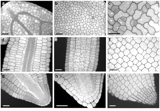
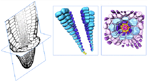
In collaboration with Jim Haseloff, we have developed new methods for fluorescent imaging of mature Arabidopsis embryos that enable their cellular architecture to be visualised without the need for labour-intensive histological sectioning. Mature embryos, which constitute an important and well-defined stage in the transition between embryogenesis and post-embryonic growth, have been particularly difficult subjects for confocal microscopy.
Using confocal microscopy, we are able to collect longitudinal optical sections through the cotyledon, hypocotyl and root of both developing and mature Arabidopsis embryos. Every cell within the embryo can be visualised with sufficient clarity and resolution to allow three-dimensional analysis of cellular architecture. The technique can be applied to the analysis of aberrant cell arrangements in Arabidopsis mutant embryos, and can be used to visualise the consequences of perturbed development such as that caused by targeted misexpression of genes in particular cells of developing meristems using GAL4 transactivation approaches. |
HOME |
LAB MEMBERS |
COORDINATION OF CELL DIVISION |
LEAF ORGANOGENESIS |
STOMATAL PATTERNING |
PhD PROJECTS |
LINKS |
Last updated 17 June 2004