MRI is a non-invasive imaging tool giving high resolution internal images to study both health and disease.
If your research sees a opportunity for MRI study please contact chym@york.ac.uk
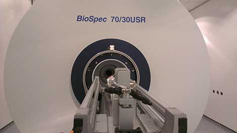
The Centre for Hyperpolarisation in MR has their own preclinical MRI facility located in the Department of Biology. This £2 million investment allows state of the art collection of MRI data to enable internal visualisation of structure with high spatial resolution. Current research focuses on the implementation of in vivo hyperpolarisation in addition to structural MRI, functional MRI and MRS (Magnetic Resonance Spectroscopy).
Equipment available:
- Range of 1H resonators and surface coils including array coils
- Wide range of X nuclei including 13C, 15N, 19F and 31P
- Dedicated anaesthetic system, animal monitoring and animal welfare apparatus
Current collaborators:
The MRI facility can be used for a range of applications including:
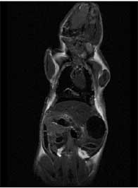
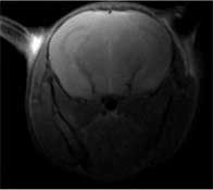
Structural
A structural MRI scan provides anatomical information about internal organs and tissue. It is used to observe structures that are normally invisible without invasive techniques. The MRI scanner can be used to examine physiological conditions associated with health in addition to spotting abnormalities and problems related to disease.
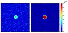
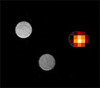
MRS
MRS (Magnetic Resonance Spectroscopy) can be used to acquire spatially resolved spectra providing information about chemical composition of a specific region. By following chemical change over time diagnostic information relating to disease can be acquired.
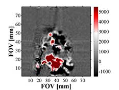
fMRI
Using fast imaging methods data can be collected which links to brain function, this is know as functional MRI (fMRI). In fMRI, changes in the distribution of the substances that carry oxygen in the blood are recorded. These changes in blood oxygenation can be related to areas of the brain which are being activated during the application of a stimulus.
Clinical MRI
As research progresses the translation of techniques from preclinical models into clinical application may be necessary. This can be made possible through links with York Neuroimaging Centre (YNiC).
-196.jpg)
Hyperpolarised MRI
Research within CHyM seeks to develop hyperpolarised methods for rapid diagnostic scanning. SABRE has the potential to revolutionise clinical MRI and related MR methods by providing a huge improvement in the sensitivity of scanners. This will ultimately produce a step change in the use and type of information available to scientists and clinicians through MRI, allowing the diagnosis, treatment and clinical monitoring of diverse neurodegenerative diseases.